
[KO Validated] Lamin A/C Rabbit pAb
Catalog Number:
ABB-A17319
- Images (9)
| Article Name: | [KO Validated] Lamin A/C Rabbit pAb |
| Biozol Catalog Number: | ABB-A17319 |
| Supplier Catalog Number: | A17319 |
| Alternative Catalog Number: | ABB-A17319-100UL, ABB-A17319-20UL |
| Manufacturer: | ABclonal |
| Host: | Rabbit |
| Category: | Antikörper |
| Application: | ELISA, IF, IHC-P, IP, WB |
| Species Reactivity: | Human |
| Immunogen: | Recombinant protein (or fragment).This information is considered to be commercially sensitive. |
| Alternative Names: | FPL, IDC, LFP, CDDC, EMD2, FPLD, HGPS, LDP1, LMN1, LMNC, MADA, PRO1, CDCD1, CMD1A, FPLD2, LMNL1, CMT2B1, LGMD1B, /C |
| The protein encoded by this gene is part of the nuclear lamina, a two-dimensional matrix of proteins located next to the inner nuclear membrane. The lamin family of proteins make up the matrix and are highly conserved in evolution. During mitosis, the lamina matrix is reversibly disassembled as the lamin proteins are phosphorylated. Lamin proteins are thought to be involved in nuclear stability, chromatin structure and gene expression. Vertebrate lamins consist of two types, A and B. Alternative splicing results in multiple transcript variants. Mutations in this gene lead to several diseases: Emery-Dreifuss muscular dystrophy, familial partial lipodystrophy, limb girdle muscular dystrophy, dilated cardiomyopathy, Charcot-Marie-Tooth disease, and Hutchinson-Gilford progeria syndrome. |
| Application Dilute: | WB,1:200 - 1:1000|IHC-P,1:50 - 1:200|IF/ICC,1:50 - 1:200|IP,0.5µg-4µg antibody for 200µg-400µg extracts of whole cells|ELISA,Recommended starting concentration is 1 µg/mL. Please optimize the concentration based on your specific assay requirements. |
| Application Notes: | Cross-Reactivity: Human,Mouse,Rat, ResearchArea: Protein phosphorylation,Signal Transduction,PI3K-Akt Signaling Pathway,Cell Biology Developmental Biology,Apoptosis,Cell Cycle,Cytoskeleton,Intermediate Filaments,Death Receptor Signaling Pathway,Stem Cells. |
|
|
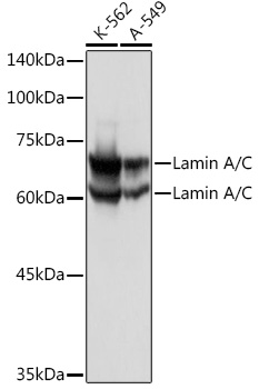
Western blot analysis of various lysates using Lamin A/C Rabbit pAb (A17319). Secondary antibody: HRP-conjugated Goat anti-Rabbit IgG (H+L) (AS014) at 1:10000 dilution. Lysates/proteins: 25µg per lane. Blocking buffer: 3% nonfat dry milk in TBST. Detection: ECL Basic Kit (RM00020). |
|
|
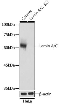
Western blot analysis of lysates from wild type (WT) and Lamin A/C knockout (KO) HeLa cells, using [KO Validated] Lamin A/C Rabbit pAb (A17319). Secondary antibody: HRP-conjugated Goat anti-Rabbit IgG (H+L) (AS014) at 1:10000 dilution. Lysates/proteins: 25µg per lane. Blocking buffer: 3% nonfat dry milk in TBST. Detection: ECL Basic Kit (RM00020). |
|
|
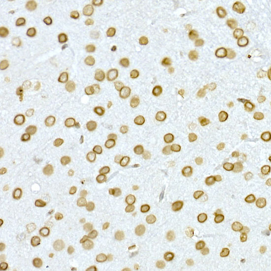
Immunohistochemistry analysis of paraffin-embedded Mouse brain using [KO Validated] Lamin A/C Rabbit pAb (A17319) at dilution of 1:50 (40x lens). High pressure antigen retrieval performed with 0.01M Citrate buffer (pH 6.0) prior to IHC staining. |
|
|
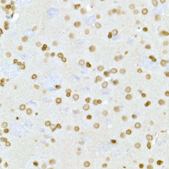
Immunohistochemistry analysis of paraffin-embedded Rat brain using [KO Validated] Lamin A/C Rabbit pAb (A17319) at dilution of 1:50 (40x lens). High pressure antigen retrieval performed with 0.01M Citrate buffer (pH 6.0) prior to IHC staining. |
|
|
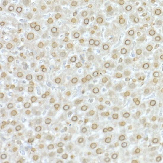
Immunohistochemistry analysis of paraffin-embedded Rat liver using [KO Validated] Lamin A/C Rabbit pAb (A17319) at dilution of 1:50 (40x lens). High pressure antigen retrieval performed with 0.01M Citrate buffer (pH 6.0) prior to IHC staining. |
|
|
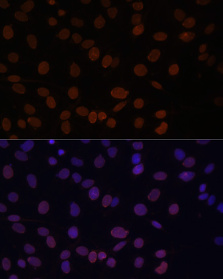
Immunofluorescence analysis of C6 cells using [KO Validated] Lamin A/C Rabbit pAb (A17319) at dilution of 1:100. Secondary antibody: Cy3-conjugated Goat anti-Rabbit IgG (H+L) (AS007) at 1:500 dilution. Blue: DAPI for nuclear staining. |
|
|
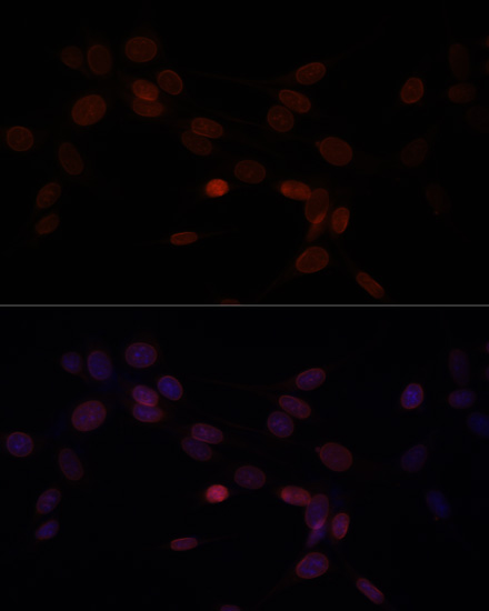
Immunofluorescence analysis of NIH-3T3 cells using [KO Validated] Lamin A/C Rabbit pAb (A17319) at dilution of 1:100. Secondary antibody: Cy3-conjugated Goat anti-Rabbit IgG (H+L) (AS007) at 1:500 dilution. Blue: DAPI for nuclear staining. |
|
|
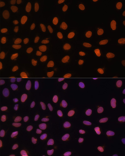
Immunofluorescence analysis of U-2 OS cells using [KO Validated] Lamin A/C Rabbit pAb (A17319) at dilution of 1:100. Secondary antibody: Cy3-conjugated Goat anti-Rabbit IgG (H+L) (AS007) at 1:500 dilution. Blue: DAPI for nuclear staining. |
|
|
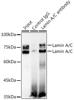
Immunoprecipitation analysis of 300 µg extracts of K-562 cells using 3 µg Lamin A/C antibody (A17319). Western blot was performed from the immunoprecipitate using Lamin A/C antibody (A17319) at a dilution of 1:1000. |
Product Guarantee and Expert Support
