
[KO Validated] APP Rabbit mAb, Unconjugated, Monoclonal
Catalog Number:
ABB-A17911
- Images (9)

| Article Name: | [KO Validated] APP Rabbit mAb, Unconjugated, Monoclonal |
| Biozol Catalog Number: | ABB-A17911 |
| Supplier Catalog Number: | A17911 |
| Alternative Catalog Number: | ABB-A17911-100UL,ABB-A17911-20UL,ABB-A17911-500UL,ABB-A17911-1000UL |
| Manufacturer: | ABclonal |
| Host: | Rabbit |
| Category: | Antikörper |
| Application: | ELISA, IF, IHC-P, IP, WB |
| Species Reactivity: | Human |
| Immunogen: | Synthetic peptide. This information is considered to be commercially sensitive. |
| Conjugation: | Unconjugated |
| Alternative Names: | AAA, AD1, PN2, ABPP, APPI, CVAP, ABETA, PN-II, preA4, CTFgamma, alpha-sAPP, PP |
| This gene encodes a cell surface receptor and transmembrane precursor protein that is cleaved by secretases to form a number of peptides. Some of these peptides are secreted and can bind to the acetyltransferase complex APBB1/TIP60 to promote transcriptional activation, while others form the protein basis of the amyloid plaques found in the brains of patients with Alzheimer disease. In addition, two of the peptides are antimicrobial peptides, having been shown to have bacteriocidal and antifungal activities. Mutations in this gene have been implicated in autosomal dominant Alzheimer disease and cerebroarterial amyloidosis (cerebral amyloid angiopathy). Multiple transcript variants encoding several different isoforms have been found for this gene. |
| Application Dilute: | WB,1:1000 - 1:6000|IP,0.5µg-4µg antibody for 200µg-400µg extracts of whole cells|IF/ICC,1:100 - 1:800|IF-P,1:100 - 1:800|IHC-P,1:2000 - 1:8000|ELISA,Recommended starting concentration is 1 µg/mL. Please optimize the concentration based on your specific as |
| Application Notes: | Cross-Reactivity: Human,Mouse,Rat. ResearchArea: Epigenetics Nuclear Signaling,Transcription Factors,Protein phosphorylation,Signal Transduction,G protein signaling,Cell Biology Developmental Biology,Apoptosis,Immunology Inflammation,Jak-Stat-IL-6 Receptor Signaling Pathway,Neuroscience,Neurodegenerative Diseases,Amyloid Plaque and Neurofibrillary Tangle Formation in Alzheimers Disease,Neurodegenerative Diseases Markers. Shipping: Ice Bag |
|
|
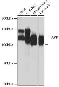
Western blot analysis of various lysates using [KO Validated] APP Rabbit mAb (A17911) at 1:1000 dilution. Secondary antibody: HRP-conjugated Goat anti-Rabbit IgG (H+L) (AS014) at 1:10000 dilution. Lysates/proteins: 25µg per lane. Blocking buffer: 3% nonfat dry milk in TBST. Detection: ECL Basic Kit (RM00020). Exposure time: 10s. |
|
|
Immunohistochemistry analysis of paraffin-embedded (5XFAD)Mouse brain and normal Mouse brain tissue using [KO Validated] APP Rabbit mAb (A17911) at a dilution of 1:2000 (40x lens). High pressure antigen retrieval performed with 0.01M Tris-EDTA Buffer (pH 9.0) prior to IHC staining. |
|
|
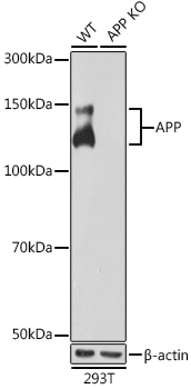
Western blot analysis of lysates from wild type (WT) and APP knockout (KO) 293T cells, using [KO Validated] APP Rabbit mAb (A17911) at 1:1000 dilution. Secondary antibody: HRP-conjugated Goat anti-Rabbit IgG (H+L) (AS014) at 1:10000 dilution. Lysates/proteins: 25µg per lane. Blocking buffer: 3% nonfat dry milk in TBST. Detection: ECL Basic Kit (RM00020). Exposure time: 10s. |
|
|
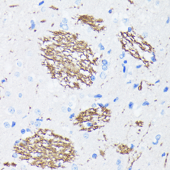
Immunohistochemistry analysis of paraffin-embedded (5XFAD)Mouse brain tissue using [KO Validated] APP Rabbit mAb (A17911) at a dilution of 1:2000 (40x lens). High pressure antigen retrieval performed with 0.01M Tris-EDTA Buffer (pH 9.0) prior to IHC staining. |
|
|
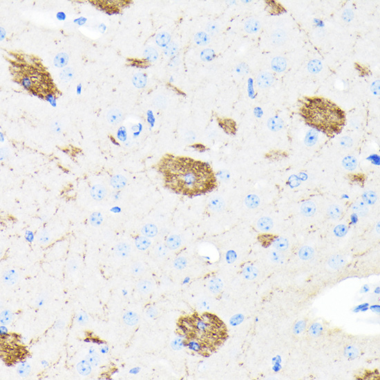
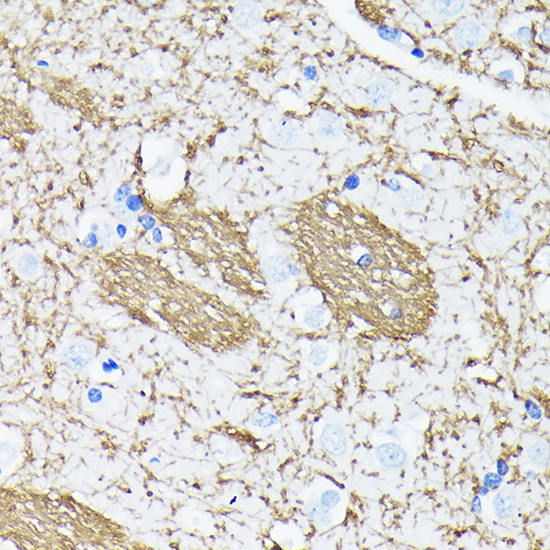
Immunohistochemistry analysis of paraffin-embedded Mouse brain tissue using [KO Validated] APP Rabbit mAb (A17911) at a dilution of 1:2000 (40x lens). High pressure antigen retrieval performed with 0.01M Tris-EDTA Buffer (pH 9.0) prior to IHC staining. |
|
|
Immunohistochemistry analysis of paraffin-embedded Rat brain tissue using [KO Validated] APP Rabbit mAb (A17911) at a dilution of 1:2000 (40x lens). High pressure antigen retrieval performed with 0.01M Tris-EDTA Buffer (pH 9.0) prior to IHC staining. |
|
|
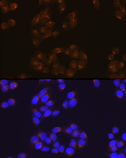
Immunofluorescence analysis of HeLa cells using APP Rabbit mAb (A17911) at dilution of 1:100 (40x lens). Secondary antibody: Cy3-conjugated Goat anti-Rabbit IgG (H+L) (AS007) at 1:500 dilution. Blue: DAPI for nuclear staining. |
|
|
Confocal imaging of paraffin-embedded 5XFAD Mouse brain and Mouse brain using [KO Validated] APP Rabbit mAb (A17911, dilution 1:200) followed by a further incubation with Cy3 Goat Anti-Rabbit IgG (H+L) (AS007, dilution 1:500) (Red). DAPI was used for nuclear staining (Blue). Objective: 40x. |
|
|
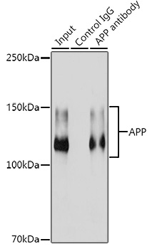
Immunoprecipitation analysis of 300 µg extracts from HeLa cells using 3 µg [KO Validated] APP Rabbit mAb (A17911). Western blot was performed from the immunoprecipitate using [KO Validated] APP Rabbit mAb (A17911) at a dilution of 1:1000. |
Product Guarantee and Expert Support
