
[KO Validated] Huntingtin Rabbit mAb, Unconjugated, Monoclonal
Catalog Number:
ABB-A19064
- Images (9)

| Article Name: | [KO Validated] Huntingtin Rabbit mAb, Unconjugated, Monoclonal |
| Biozol Catalog Number: | ABB-A19064 |
| Supplier Catalog Number: | A19064 |
| Alternative Catalog Number: | ABB-A19064-20UL,ABB-A19064-100UL |
| Manufacturer: | ABclonal |
| Host: | Rabbit |
| Category: | Antikörper |
| Application: | ELISA, IF, WB |
| Species Reactivity: | Human |
| Immunogen: | Synthetic peptide. This information is considered to be commercially sensitive. |
| Conjugation: | Unconjugated |
| Alternative Names: | HD, IT15, LOMARS, Huntingtin |
| Huntingtin is a disease gene linked to Huntingtons disease, a neurodegenerative disorder characterized by loss of striatal neurons. This is thought to be caused by an expanded, unstable trinucleotide repeat in the huntingtin gene, which translates as a polyglutamine repeat in the protein product. A fairly broad range of trinucleotide repeats (9-35) has been identified in normal controls, and repeat numbers in excess of 40 have been described as pathological. The huntingtin locus is large, spanning 180 kb and consisting of 67 exons. The huntingtin gene is widely expressed and is required for normal development. It is expressed as 2 alternatively polyadenylated forms displaying different relative abundance in various fetal and adult tissues. The larger transcript is approximately 13.7 kb and is expressed predominantly in adult and fetal brain whereas the smaller transcript of approximately 10.3 kb is more widely expressed. The genetic defect leading to Huntingtons disease may not necessarily eliminate transcription, but may confer a new property on the mRNA or alter the function of the protein. One candidate is the huntingtin-associated protein-1, highly expressed in brain, which has increased affinity for huntingtin protein with expanded polyglutamine repeats. This gene contains an upstream open reading frame in the 5 UTR that inhibits expression of the huntingtin gene product through translational repression. |
| Application Dilute: | WB,1:500 - 1:1000|IF-F,1:100 - 1:200|ELISA,Recommended starting concentration is 1 µg/mL. Please optimize the concentration based on your specific assay requirements. |
| Application Notes: | Cross-Reactivity: Human,Mouse,Rat. ResearchArea: Signal Transduction,PI3K-Akt Signaling Pathway,Cell Biology Developmental Biology,Apoptosis,Endocrine Metabolism,Mitochondrial metabolism,Neuroscience,Neurodegenerative Diseases,Neurodegenerative Diseases Markers,Other Neurological disorders. Shipping: Ice Bag |
|
|
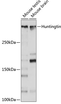
Western blot analysis of various lysates using [KO Validated] Huntingtin Rabbit mAb (A19064) at 1:1000 dilution. Secondary antibody: HRP-conjugated Goat anti-Rabbit IgG (H+L) (AS014) at 1:10000 dilution. Lysates/proteins: 25µg per lane. Blocking buffer: 3% nonfat dry milk in TBST. Detection: ECL Basic Kit (RM00020). Exposure time: 10s. |
|
|
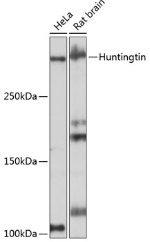
Western blot analysis of various lysates using [KO Validated] Huntingtin Rabbit mAb at 1:1000 dilution. Secondary antibody: HRP-conjugated Goat anti-Rabbit IgG (H+L) (AS014) at 1:10000 dilution. Lysates/proteins: 25µg per lane. Blocking buffer: 3% nonfat dry milk in TBST. Detection: ECL Basic Kit (RM00020). Exposure time: 3min. |
|
|
Confocal imaging of frozen sections Mouse brain tissue using [KO Validated] Huntingtin Rabbit mAb (A19064, dilution 1:100) followed by a further incubation with Cy3-conjugated Goat Anti-Rabbit IgG (H+L) (AS007, dilution 1:500) (Red). DAPI was used for nuclear staining (Blue). Objective: 40x. |
|
|
Confocal imaging of frozen sections Rat brain tissue using [KO Validated] Huntingtin Rabbit mAb (A19064, dilution 1:100) followed by a further incubation with Cy3-conjugated Goat Anti-Rabbit IgG (H+L) (AS007, dilution 1:500) (Red). DAPI was used for nuclear staining (Blue). Objective: 40x. |
|
|
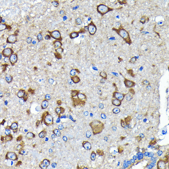
Immunohistochemistry analysis of paraffin-embeddedRat brain tissue using[KO Validated] Huntingtin Rabbit mAb(A19064) at a dilution of 1:200 (40x lens).High pressure antigen retrieval was performed with 0.01 M citrate buffer (pH 6.0) prior to IHC staining. |
|
|
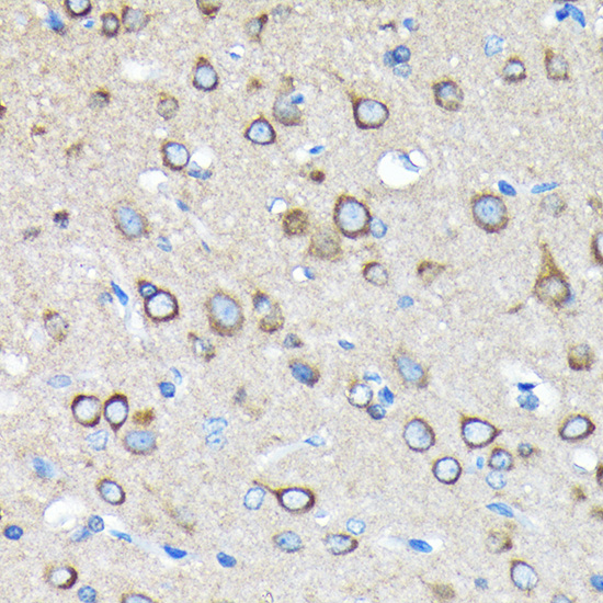
Immunohistochemistry analysis of paraffin-embeddedMouse brain tissue using[KO Validated] Huntingtin Rabbit mAb(A19064) at a dilution of 1:200 (40x lens).High pressure antigen retrieval was performed with 0.01M citrate buffer (pH 6.0) prior to IHC staining. |
|
|
Immunohistochemistry analysis of paraffin-embeddedHuman brain tissue using[KO Validated]Huntingtin Rabbit mAb(A19064) at a dilution of 1:200 (40x lens).High pressure antigen retrieval was performed with 0.01M citrate buffer (pH 6.0) prior to IHC staining. |
|
|
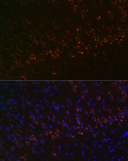
Immunofluorescence analysis of Mouse brain tissue using [KO Validated] Huntingtin Rabbit mAb (A19064) at a dilution of 1:100 (40x lens). Secondary antibody: Cy3-conjugated Goat anti-Rabbit IgG (H+L)(AS007) at 1:500 dilution. Blue: DAPI for nuclear staining. Microwave antigen retrieval performed with 0.01M Citrate Buffer(pH 6.0) prior to IF staining. |
|
|
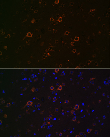
Immunofluorescence analysis of Rat brain tissue using [KO Validated] Huntingtin Rabbit mAb (A19064) at a dilution of 1:100 (40x lens). Secondary antibody: Cy3-conjugated Goat anti-Rabbit IgG (H+L)(AS007) at 1:500 dilution. Blue: DAPI for nuclear staining. Microwave antigen retrieval performed with 0.01M Citrate Buffer(pH 6.0) prior to IF staining. |
Product Guarantee and Expert Support
