
[KO Validated] SUMO1 Rabbit mAb, Unconjugated, Monoclonal
Catalog Number:
ABB-A19121
- Images (9)

| Article Name: | [KO Validated] SUMO1 Rabbit mAb, Unconjugated, Monoclonal |
| Biozol Catalog Number: | ABB-A19121 |
| Supplier Catalog Number: | A19121 |
| Alternative Catalog Number: | ABB-A19121-100UL,ABB-A19121-20UL,ABB-A19121-500UL,ABB-A19121-1000UL |
| Manufacturer: | ABclonal |
| Host: | Rabbit |
| Category: | Antikörper |
| Application: | ELISA, IF, IHC-P, WB |
| Species Reactivity: | Human |
| Immunogen: | Synthetic peptide. This information is considered to be commercially sensitive. |
| Conjugation: | Unconjugated |
| Alternative Names: | DAP1, GMP1, PIC1, SMT3, UBL1, OFC10, SENP2, SMT3C, SMT3H3, O1 |
| This gene encodes a protein that is a member of the SUMO (small ubiquitin-like modifier) protein family. It functions in a manner similar to ubiquitin in that it is bound to target proteins as part of a post-translational modification system. However, unlike ubiquitin which targets proteins for degradation, this protein is involved in a variety of cellular processes, such as nuclear transport, transcriptional regulation, apoptosis, and protein stability. It is not active until the last four amino acids of the carboxy-terminus have been cleaved off. Several pseudogenes have been reported for this gene. Alternate transcriptional splice variants encoding different isoforms have been characterized. |
| Application Dilute: | WB,1:1000 - 1:2000|IHC-P,1:2000 - 1:10000|IF/ICC,1:200 - 1:400|ELISA,Recommended starting concentration is 1 µg/mL. Please optimize the concentration based on your specific assay requirements. |
| Application Notes: | Cross-Reactivity: Human,Mouse,Rat. ResearchArea: Epigenetics Nuclear Signaling,RNA Binding,Cell Biology Developmental Biology,Autophagy,Ubiquitin,Endocrine Metabolism,Mitochondrial metabolism,Immunology Inflammation,Jak-Stat-IL-6 Receptor Signaling Pathway,NF-kB Signaling Pathway,Cardiovascular,Heart. Shipping: Ice Bag |
|
|
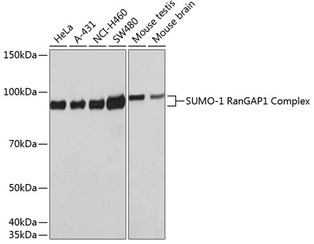
Western blot analysis of various lysates using [KO Validated] SUMO1 Rabbit mAb (A19121) at 1:1000 dilution. Secondary antibody: HRP-conjugated Goat anti-Rabbit IgG (H+L) (AS014) at 1:10000 dilution. Lysates/proteins: 25µg per lane. Blocking buffer: 3% nonfat dry milk in TBST. Detection: ECL Basic Kit (RM00020). Exposure time: 10s. |
|
|
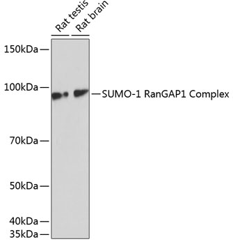
Western blot analysis of various lysates using [KO Validated] SUMO1 Rabbit mAb (A19121) at 1:1000 dilution. Secondary antibody: HRP-conjugated Goat anti-Rabbit IgG (H+L) (AS014) at 1:10000 dilution. Lysates/proteins: 25µg per lane. Blocking buffer: 3% nonfat dry milk in TBST. Detection: ECL Basic Kit (RM00020). Exposure time: 1min. |
|
|
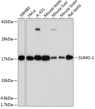
Western blot analysis of various lysates using [KO Validated] SUMO1 Rabbit mAb (A19121) at 1:1000 dilution. Secondary antibody: HRP-conjugated Goat anti-Rabbit IgG (H+L) (AS014) at 1:10000 dilution. Lysates/proteins: 25µg per lane. Blocking buffer: 3% nonfat dry milk in TBST. Detection: ECL Basic Kit (RM00020). Exposure time: 60s. |
|
|
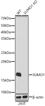
Western blot analysis of lysates from wild type(WT) and SUMO1 knockout (KO) 293T cells, using [KO Validated] SUMO1 Rabbit mAb (A19121) at 1:1000 dilution. Secondary antibody: HRP-conjugated Goat anti-Rabbit IgG (H+L) (AS014) at 1:10000 dilution. Lysates/proteins: 25µg per lane. Blocking buffer: 3% nonfat dry milk in TBST. Detection: ECL Basic Kit (RM00020). Exposure time: 10s. |
|
|
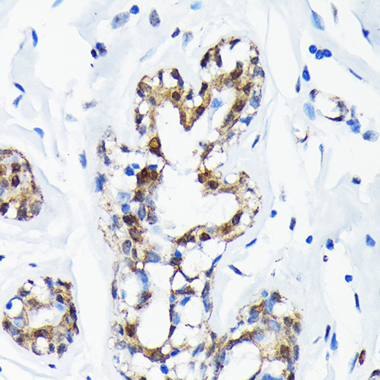
Immunohistochemistry analysis of paraffin-embedded Human esophagus tissue using [KO Validated] SUMO1 Rabbit mAb (A19121) at a dilution of 1:5000 (40x lens). High pressure antigen retrieval performed with 0.01M Tris-EDTA Buffer (pH 9.0) prior to IHC staining. |
|
|
Immunohistochemistry analysis of paraffin-embedded Human tonsil tissue using [KO Validated] SUMO1 Rabbit mAb (A19121) at a dilution of 1:5000 (40x lens). High pressure antigen retrieval performed with 0.01M Tris-EDTA Buffer (pH 9.0) prior to IHC staining. |
|
|
Immunohistochemistry analysis of paraffin-embedded Mouse brain tissue using [KO Validated] SUMO1 Rabbit mAb (A19121) at a dilution of 1:5000 (40x lens). High pressure antigen retrieval performed with 0.01M Tris-EDTA Buffer (pH 9.0) prior to IHC staining. |
|
|
Immunohistochemistry analysis of paraffin-embedded Rat spleen tissue using [KO Validated] SUMO1 Rabbit mAb (A19121) at a dilution of 1:5000 (40x lens). High pressure antigen retrieval performed with 0.01M Tris-EDTA Buffer (pH 9.0) prior to IHC staining. |
|
|
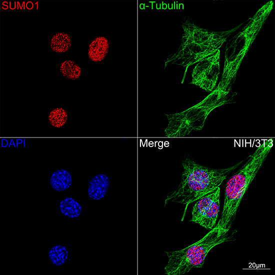
Confocal imaging of NIH/3T3 cells using [KO Validated] SUMO1 Rabbit mAb (A19121,dilution 1:100)(Red). The cells were counterstained with alpha-Tubulin Mouse mAb (AC012,dilution 1:400) (Green). DAPI was used for nuclear staining (blue). Objective: 100x. |
Product Guarantee and Expert Support
