
CD27 Rabbit pAb, Unconjugated, Polyclonal
Catalog Number:
ABB-A1945
- Images (9)
| Article Name: | CD27 Rabbit pAb, Unconjugated, Polyclonal |
| Biozol Catalog Number: | ABB-A1945 |
| Supplier Catalog Number: | A1945 |
| Alternative Catalog Number: | ABB-A1945-100UL,ABB-A1945-20UL,ABB-A1945-500UL,ABB-A1945-1000UL |
| Manufacturer: | ABclonal |
| Host: | Rabbit |
| Category: | Antikörper |
| Application: | ELISA, IF, IHC-P, WB |
| Species Reactivity: | Human |
| Immunogen: | Recombinant protein (or fragment).This information is considered to be commercially sensitive. |
| Conjugation: | Unconjugated |
| Alternative Names: | T14, S152, Tp55, TNFRSF7, S152. LPFS2, CD27 |
| The protein encoded by this gene is a member of the TNF-receptor superfamily. This receptor is required for generation and long-term maintenance of T cell immunity. It binds to ligand CD70, and plays a key role in regulating B-cell activation and immunoglobulin synthesis. This receptor transduces signals that lead to the activation of NF-kappaB and MAPK8/JNK. Adaptor proteins TRAF2 and TRAF5 have been shown to mediate the signaling process of this receptor. CD27-binding protein (SIVA), a proapoptotic protein, can bind to this receptor and is thought to play an important role in the apoptosis induced by this receptor. |
| Clonality: | Polyclonal |
| Molecular Weight: | 29kDa |
| NCBI: | 939 |
| UniProt: | P26842 |
| Purity: | Affinity purification |
| Sequence: | ATPAPKSCPERHYWAQGKLCCQMCEPGTFLVKDCDQHRKAAQCDPCIPGVSFSPDHHTRPHCESCRHCNSGLLVRNCTITANAECACRNGWQCRDKECTECDPLPNPSLTARSSQALSPHPQPTHLPYVSEMLEARTAGHMQTLADFRQLPARTLSTHWPPQRSLCSSDFIR |
| Target: | CD27 |
| Antibody Type: | Primary Antibody |
| Application Dilute: | WB,1:500 - 1:1000|IF/ICC,1:50 - 1:200|IF-P,1:50 - 1:200|IHC-P,1:50 - 1:200|ELISA,Recommended starting concentration is 1 µg/mL. Please optimize the concentration based on your specific assay requirements. |
| Application Notes: | Cross-Reactivity: Human,Mouse,Rat. ResearchArea: Cancer,Tumor immunology,Cell Biology Developmental Biology,Apoptosis,Immunology Inflammation,CDs,Cytokines,Cell Intrinsic Innate Immunity Signaling Pathway,Stem Cells,Hematopoietic Progenitors. Shipping: Ice Bag |
|
|
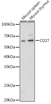
Western blot analysis of various lysates using CD27 Rabbit pAb (A1945) at 1:1000 dilution. Secondary antibody: HRP-conjugated Goat anti-Rabbit IgG (H+L) (AS014) at 1:10000 dilution. Lysates/proteins: 25µg per lane. Blocking buffer: 3% nonfat dry milk in TBST. Detection: ECL Basic Kit (RM00020). Exposure time: 10s. |
|
|
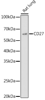
Western blot analysis of lysates from Rat lung, using CD27 Rabbit pAb (A1945) at 1:1000 dilution. Secondary antibody: HRP-conjugated Goat anti-Rabbit IgG (H+L) (AS014) at 1:10000 dilution. Lysates/proteins: 25µg per lane. Blocking buffer: 3% nonfat dry milk in TBST. Detection: ECL Basic Kit (RM00020). Exposure time: 90s. |
|
|
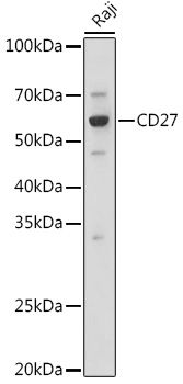
Western blot analysis of lysates from Raji cells, using CD27 Rabbit pAb (A1945) at 1:1000 dilution. Secondary antibody: HRP-conjugated Goat anti-Rabbit IgG (H+L) (AS014) at 1:10000 dilution. Lysates/proteins: 25µg per lane. Blocking buffer: 3% nonfat dry milk in TBST. Detection: ECL Basic Kit (RM00020). Exposure time: 3s. |
|
|
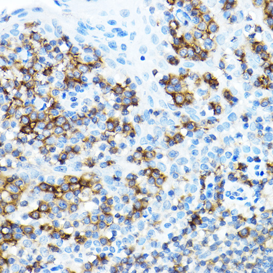
Immunohistochemistry analysis of paraffin-embedded Human tonsil using CD27 Rabbit pAb (A1945) at dilution of 1:100 (40x lens). High pressure antigen retrieval performed with 0.01M Citrate buffer (pH 6.0) prior to IHC staining. |
|
|
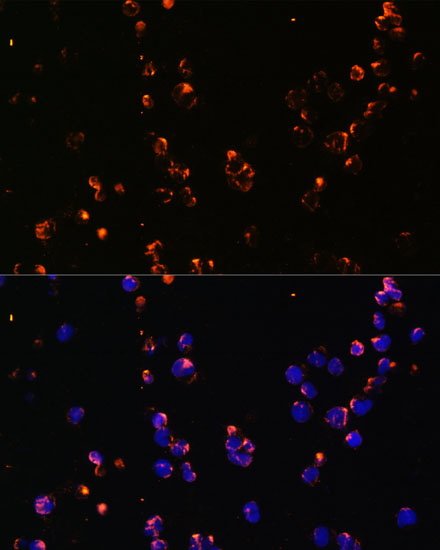
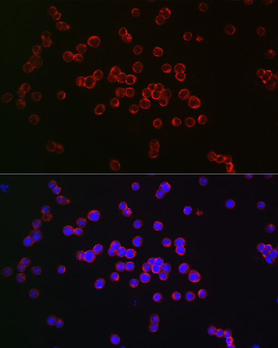
Immunofluorescence analysis of Jurkat cells using CD27 Rabbit pAb (A1945) at dilution of 1:100. Secondary antibody: Cy3-conjugated Goat anti-Rabbit IgG (H+L) (AS007) at 1:500 dilution. Blue: DAPI for nuclear staining. |
|
|
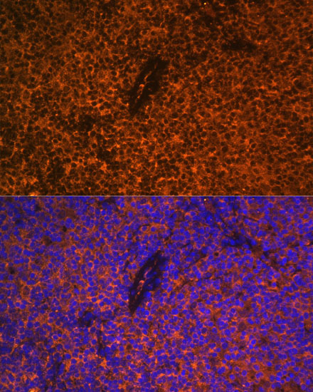
Immunofluorescence analysis of paraffin-embedded rat thymus using CD27 Rabbit pAb (A1945) at dilution of 1:100. Secondary antibody: Cy3-conjugated Goat anti-Rabbit IgG (H+L) (AS007) at 1:500 dilution. Blue: DAPI for nuclear staining. |
|
|
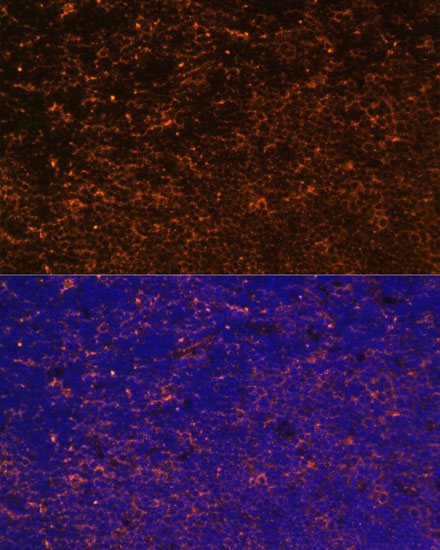
Immunofluorescence analysis of paraffin-embedded mouse thymus using CD27 Rabbit pAb (A1945) at dilution of 1:100. Secondary antibody: Cy3-conjugated Goat anti-Rabbit IgG (H+L) (AS007) at 1:500 dilution. Blue: DAPI for nuclear staining. |
|
|
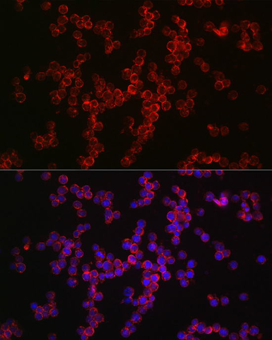
Immunofluorescence analysis of Jurkat cells using CD27 Rabbit pAb (A1945) at dilution of 1:100 (40x lens). Secondary antibody: Cy3-conjugated Goat anti-Rabbit IgG (H+L) (AS007) at 1:500 dilution. Blue: DAPI for nuclear staining. |
|
|
Immunofluorescence analysis of Raji cells using CD27 Rabbit pAb (A1945) at dilution of 1:100 (40x lens). Secondary antibody: Cy3-conjugated Goat anti-Rabbit IgG (H+L) (AS007) at 1:500 dilution. Blue: DAPI for nuclear staining. |
Product Guarantee and Expert Support
