
AIF Rabbit mAb, Unconjugated, Monoclonal
Catalog Number:
ABB-A19536
- Images (9)

| Article Name: | AIF Rabbit mAb, Unconjugated, Monoclonal |
| Biozol Catalog Number: | ABB-A19536 |
| Supplier Catalog Number: | A19536 |
| Alternative Catalog Number: | ABB-A19536-20UL,ABB-A19536-100UL |
| Manufacturer: | ABclonal |
| Host: | Rabbit |
| Category: | Antikörper |
| Application: | ELISA, IHC-P, WB |
| Species Reactivity: | Human |
| Immunogen: | Synthetic peptide. This information is considered to be commercially sensitive. |
| Conjugation: | Unconjugated |
| Alternative Names: | AIF, AUNX1, CMT2D, CMTX4, COWCK, DFNX5, NADMR, NAMSD, PDCD8, COXPD6, SEMDHL |
| This gene encodes a flavoprotein essential for nuclear disassembly in apoptotic cells, and it is found in the mitochondrial intermembrane space in healthy cells. Induction of apoptosis results in the translocation of this protein to the nucleus where it affects chromosome condensation and fragmentation. In addition, this gene product induces mitochondria to release the apoptogenic proteins cytochrome c and caspase-9. Mutations in this gene cause combined oxidative phosphorylation deficiency 6 (COXPD6), a severe mitochondrial encephalomyopathy, as well as Cowchock syndrome, also known as X-linked recessive Charcot-Marie-Tooth disease-4 (CMTX-4), a disorder resulting in neuropathy, and axonal and motor-sensory defects with deafness and cognitive disability. Alternative splicing results in multiple transcript variants. A related pseudogene has been identified on chromosome 10. |
| Application Dilute: | WB,1:1000 - 1:4000|IHC-P,1:200 - 1:800|ELISA,Recommended starting concentration is 1 µg/mL. Please optimize the concentration based on your specific assay requirements. |
| Application Notes: | Cross-Reactivity: Human,Mouse,Rat. ResearchArea: Cell Biology Developmental Biology,Apoptosis,Mitochondrial Control of Apoptosis. Shipping: Ice Bag |
|
|
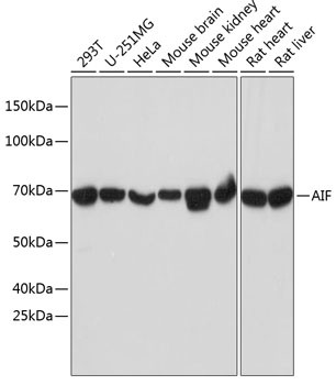
Western blot analysis of various lysates using AIF Rabbit mAb (A19536) at 1:1000 dilution. Secondary antibody: HRP-conjugated Goat anti-Rabbit IgG (H+L) (AS014) at 1:10000 dilution. Lysates/proteins: 25µg per lane. Blocking buffer: 3% nonfat dry milk in TBST. Detection: ECL Basic Kit (RM00020). Exposure time: 30s. |
|
|
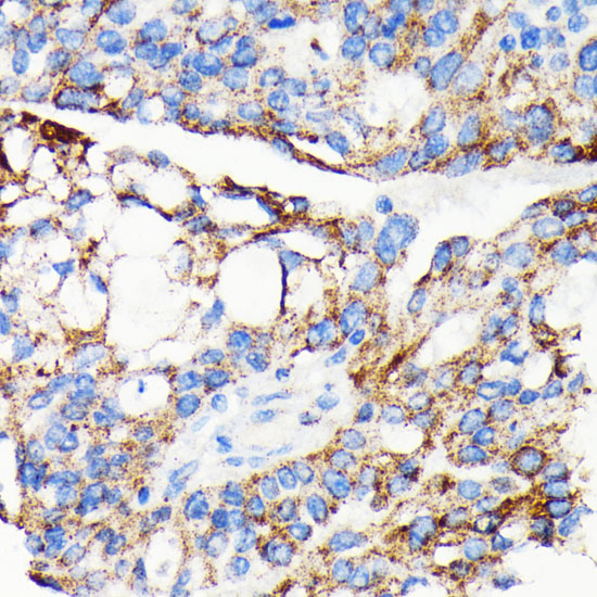
Immunohistochemistry analysis of paraffin-embedded Human colon carcinoma tissue using AIF Rabbit mAb (A19536) at a dilution of 1:500 (40x lens). High pressure antigen retrieval performed with 0.01M Tris-EDTA Buffer (pH 9.0) prior to IHC staining. |
|
|
Immunohistochemistry analysis of paraffin-embedded Human kidney tissue using AIF Rabbit mAb (A19536) at a dilution of 1:500 (40x lens). High pressure antigen retrieval performed with 0.01M Tris-EDTA Buffer (pH 9.0) prior to IHC staining. |
|
|
Immunohistochemistry analysis of paraffin-embedded Human liver tissue using AIF Rabbit mAb (A19536) at a dilution of 1:500 (40x lens). High pressure antigen retrieval performed with 0.01M Tris-EDTA Buffer (pH 9.0) prior to IHC staining. |
|
|
Immunohistochemistry analysis of paraffin-embedded Human lung squamous carcinoma tissue using AIF Rabbit mAb (A19536) at a dilution of 1:500 (40x lens). High pressure antigen retrieval performed with 0.01M Tris-EDTA Buffer (pH 9.0) prior to IHC staining. |
|
|
Immunohistochemistry analysis of paraffin-embedded Human pancreas tissue using AIF Rabbit mAb (A19536) at a dilution of 1:500 (40x lens). High pressure antigen retrieval performed with 0.01M Tris-EDTA Buffer (pH 9.0) prior to IHC staining. |
|
|
Immunohistochemistry analysis of paraffin-embedded Mouse liver tissue using AIF Rabbit mAb (A19536) at a dilution of 1:500 (40x lens). High pressure antigen retrieval performed with 0.01M Tris-EDTA Buffer (pH 9.0) prior to IHC staining. |
|
|
Immunohistochemistry analysis of paraffin-embedded Rat colon tissue using AIF Rabbit mAb (A19536) at a dilution of 1:500 (40x lens). High pressure antigen retrieval performed with 0.01M Tris-EDTA Buffer (pH 9.0) prior to IHC staining. |
|
|
Immunohistochemistry analysis of paraffin-embedded Rat testis tissue using AIF Rabbit mAb (A19536) at a dilution of 1:500 (40x lens). High pressure antigen retrieval performed with 0.01M Tris-EDTA Buffer (pH 9.0) prior to IHC staining. |
Product Guarantee and Expert Support
