
[KO Validated] HuR/ELAVL1 Rabbit mAb, Unconjugated, Monoclonal
Catalog Number:
ABB-A19622
- Images (9)

| Article Name: | [KO Validated] HuR/ELAVL1 Rabbit mAb, Unconjugated, Monoclonal |
| Biozol Catalog Number: | ABB-A19622 |
| Supplier Catalog Number: | A19622 |
| Alternative Catalog Number: | ABB-A19622-100UL,ABB-A19622-20UL,ABB-A19622-500UL,ABB-A19622-1000UL |
| Manufacturer: | ABclonal |
| Host: | Rabbit |
| Category: | Antikörper |
| Application: | ELISA, IF, IHC-P, IP, WB |
| Species Reactivity: | Human |
| Immunogen: | Synthetic peptide. This information is considered to be commercially sensitive. |
| Conjugation: | Unconjugated |
| Alternative Names: | HUR, Hua, MelG, ELAV1, L1 |
| The protein encoded by this gene is a member of the ELAVL family of RNA-binding proteins that contain several RNA recognition motifs, and selectively bind AU-rich elements (AREs) found in the 3 untranslated regions of mRNAs. AREs signal degradation of mRNAs as a means to regulate gene expression, thus by binding AREs, the ELAVL family of proteins play a role in stabilizing ARE-containing mRNAs. This gene has been implicated in a variety of biological processes and has been linked to a number of diseases, including cancer. It is highly expressed in many cancers, and could be potentially useful in cancer diagnosis, prognosis, and therapy. |
| Application Dilute: | WB,1:1000 - 1:6000|IHC-P,1:200 - 1:2000|IF/ICC,1:100 - 1:1000|IP,0.5µg-4µg antibody for 200µg-400µg extracts of whole cells|ELISA,Recommended starting concentration is 1 µg/mL. Please optimize the concentration based on your specific assay requirements. |
| Application Notes: | Cross-Reactivity: Human,Mouse,Rat. ResearchArea: Epigenetics Nuclear Signaling,RNA Binding,Endocrine Metabolism,AMPK Signaling Pathway. Shipping: Ice Bag |
|
|
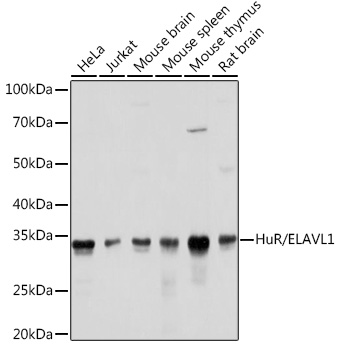
Western blot analysis of various lysates using [KO Validated] HuR/ELAVL1 Rabbit mAb (A19622) at 1:1000 dilution. Secondary antibody: HRP-conjugated Goat anti-Rabbit IgG (H+L) (AS014) at 1:10000 dilution. Lysates/proteins: 25µg per lane. Blocking buffer: 3% nonfat dry milk in TBST. Detection: ECL Basic Kit (RM00020). Exposure time: 1s. |
|
|
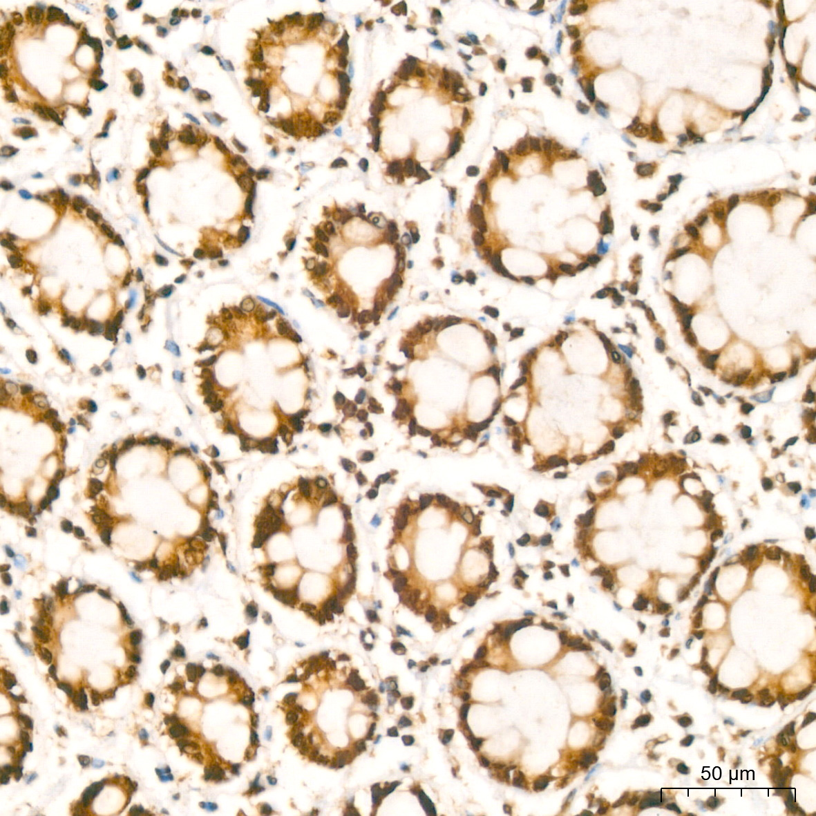
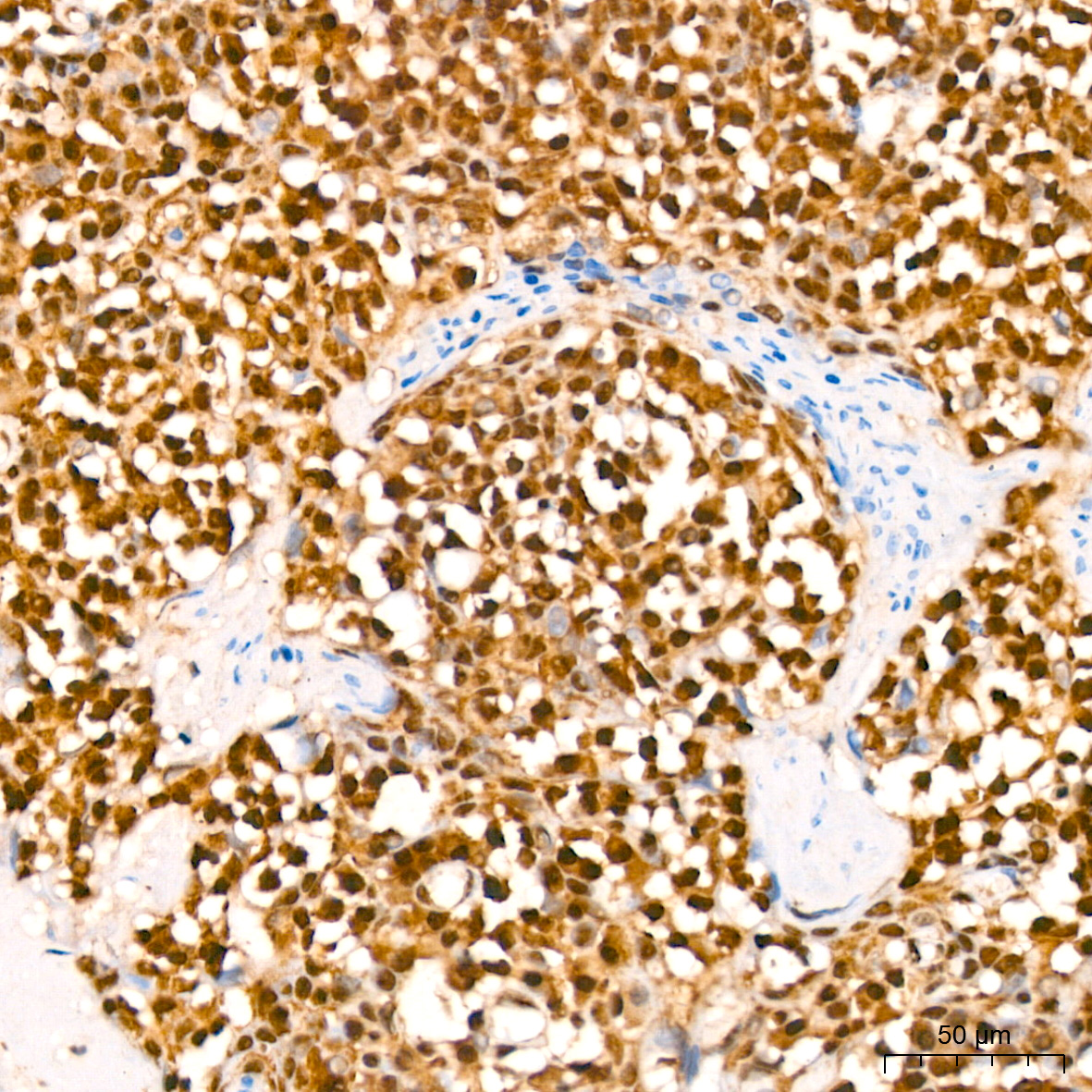
Immunohistochemistry analysis of paraffin-embedded Human colon carcinoma using [KO Validated] HuR/ELAVL1 Rabbit mAb (A19622) at dilution of 1:200 (40x lens). High pressure antigen retrieval performed with 0.01M Citrate buffer (pH 6.0) prior to IHC staining. |
|
|
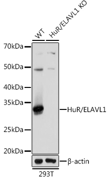
Western blot analysis of lysates from wild type (WT) and HuR/ELAVL1 knockout (KO) 293T cells, using [KO Validated] HuR/ELAVL1 Rabbit mAb (A19622) at 1:1000 dilution. Secondary antibody: HRP-conjugated Goat anti-Rabbit IgG (H+L) (AS014) at 1:10000 dilution. Lysates/proteins: 25µg per lane. Blocking buffer: 3% nonfat dry milk in TBST. Detection: ECL Basic Kit (RM00020). Exposure time: 1s. |
|
|
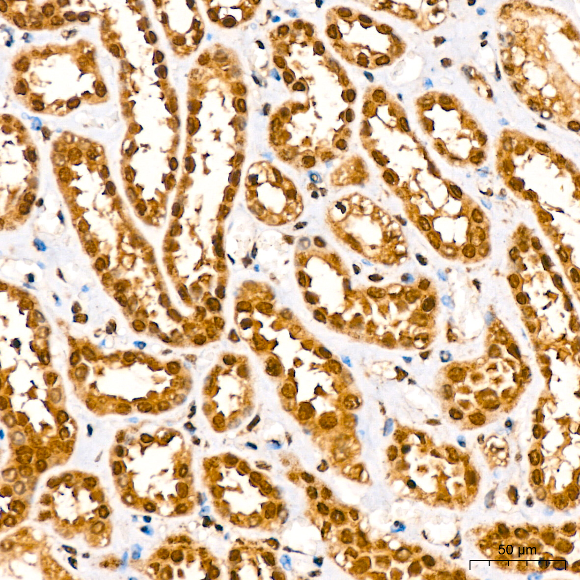
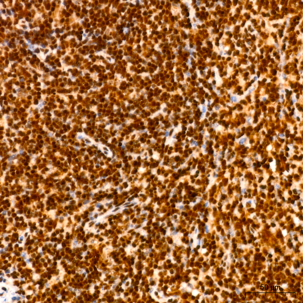
Immunohistochemistry analysis of paraffin-embedded Human kidney using [KO Validated] HuR/ELAVL1 Rabbit mAb (A19622) at dilution of 1:200 (40x lens). High pressure antigen retrieval performed with 0.01M Citrate buffer (pH 6.0) prior to IHC staining. |
|
|
Immunohistochemistry analysis of paraffin-embedded Human tonsil using [KO Validated] HuR/ELAVL1 Rabbit mAb (A19622) at dilution of 1:200 (40x lens). High pressure antigen retrieval performed with 0.01M Citrate buffer (pH 6.0) prior to IHC staining. |
|
|
Immunohistochemistry analysis of paraffin-embedded Mouse spleen using [KO Validated] HuR/ELAVL1 Rabbit mAb (A19622) at dilution of 1:200 (40x lens). High pressure antigen retrieval performed with 0.01M Citrate buffer (pH 6.0) prior to IHC staining. |
|
|
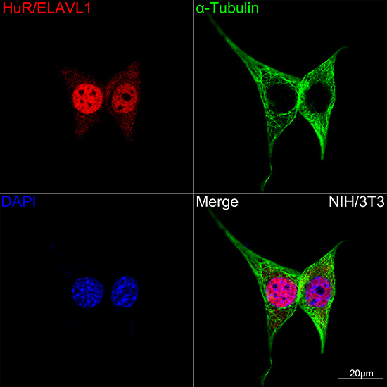
Confocal imaging of NIH/3T3 cells using [KO Validated] HuR/ELAVL1 Rabbit mAb (A19622,dilution 1:100)(Red). The cells were counterstained with alpha-Tubulin Mouse mAb (AC012,dilution 1:400) (Green). DAPI was used for nuclear staining (blue). Objective: 100x. |
|
|
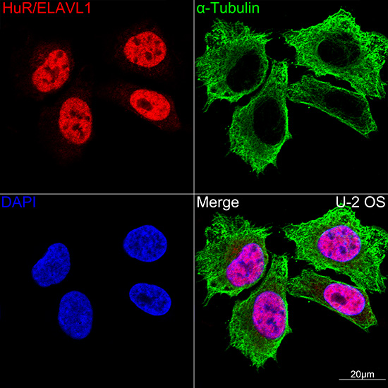
Confocal imaging of U-2 OS cells using [KO Validated] HuR/ELAVL1 Rabbit mAb (A19622,dilution 1:100)(Red). The cells were counterstained with alpha-Tubulin Mouse mAb (AC012,dilution 1:400) (Green). DAPI was used for nuclear staining (blue). Objective: 100x. |
|
|
Immunoprecipitation analysis of 300 µg extracts of 293T cells using 3 µg HuR/ELAVL1 antibody (A19622). Western blot was performed from the immunoprecipitate using HuR/ELAVL1 antibody (A19622) at a dilution of 1:1000. |
Product Guarantee and Expert Support
