
TMPRSS2 Rabbit pAb, Unconjugated, Polyclonal
Catalog Number:
ABB-A1979
- Images (6)


| Article Name: | TMPRSS2 Rabbit pAb, Unconjugated, Polyclonal |
| Biozol Catalog Number: | ABB-A1979 |
| Supplier Catalog Number: | A1979 |
| Alternative Catalog Number: | ABB-A1979-100UL,ABB-A1979-20UL,ABB-A1979-500UL,ABB-A1979-1000UL |
| Manufacturer: | ABclonal |
| Host: | Rabbit |
| Category: | Antikörper |
| Application: | ELISA, IF, IHC-P, WB |
| Species Reactivity: | Human |
| Immunogen: | Synthetic peptide. This information is considered to be commercially sensitive. |
| Conjugation: | Unconjugated |
| Alternative Names: | PRSS10, TMPRSS2 |
| This gene encodes a protein that belongs to the serine protease family. The encoded protein contains a type II transmembrane domain, a receptor class A domain, a scavenger receptor cysteine-rich domain and a protease domain. Serine proteases are known to be involved in many physiological and pathological processes. This gene was demonstrated to be up-regulated by androgenic hormones in prostate cancer cells and down-regulated in androgen-independent prostate cancer tissue. The protease domain of this protein is thought to be cleaved and secreted into cell media after autocleavage. This protein also facilitates entry of viruses into host cells by proteolytically cleaving and activating viral envelope glycoproteins. Viruses found to use this protein for cell entry include Influenza virus and the human coronaviruses HCoV-229E, MERS-CoV, SARS-CoV and SARS-CoV-2 (COVID-19 virus). Alternatively spliced transcript variants encoding different isoforms have been found for this gene. |
| Application Dilute: | WB,1:500 - 1:1000|IHC-P,1:50 - 1:200|IF/ICC,1:50 - 1:200|ELISA,Recommended starting concentration is 1 µg/mL. Please optimize the concentration based on your specific assay requirements. |
| Application Notes: | Cross-Reactivity: Human,Mouse,Rat. ResearchArea: Cancer,Tumor biomarkers,Cell Biology Developmental Biology,Ubiquitin,Neuroscience,Neurodegenerative Diseases. Shipping: Ice Bag |
|
|
Immunohistochemistry analysis of paraffin-embedded Human breast cancer using TMPRSS2 Rabbit pAb (A1979) at dilution of 1:50 (40x lens). High pressure antigen retrieval performed with 0.01M Citrate buffer (pH 6.0) prior to IHC staining. |
|
|
Immunohistochemistry analysis of paraffin-embedded Human esophageal cancer using TMPRSS2 Rabbit pAb (A1979) at dilution of 1:50 (40x lens). High pressure antigen retrieval performed with 0.01M Citrate buffer (pH 6.0) prior to IHC staining. |
|
|
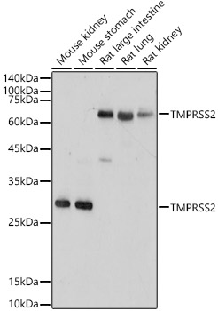
Western blot analysis of various lysates using TMPRSS2 Rabbit pAb (A1979) at 1:1000 dilution. Secondary antibody: HRP-conjugated Goat anti-Rabbit IgG (H+L) (AS014) at 1:10000 dilution. Lysates/proteins: 25µg per lane. Blocking buffer: 3% nonfat dry milk in TBST. Detection: ECL Enhanced Kit (RM00021). Exposure time: 180s. |
|
|
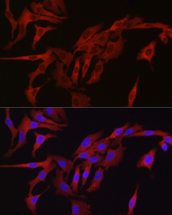
Immunofluorescence analysis of A-549 cells using TMPRSS2 Rabbit pAb (A1979) at dilution of 1:50 (40x lens). Secondary antibody: Cy3-conjugated Goat anti-Rabbit IgG (H+L) (AS007) at 1:500 dilution. Blue: DAPI for nuclear staining. |
|
|
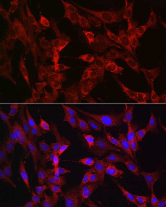
Immunofluorescence analysis of C6 cells using TMPRSS2 Rabbit pAb (A1979) at dilution of 1:50 (40x lens). Secondary antibody: Cy3-conjugated Goat anti-Rabbit IgG (H+L) (AS007) at 1:500 dilution. Blue: DAPI for nuclear staining. |
|
|
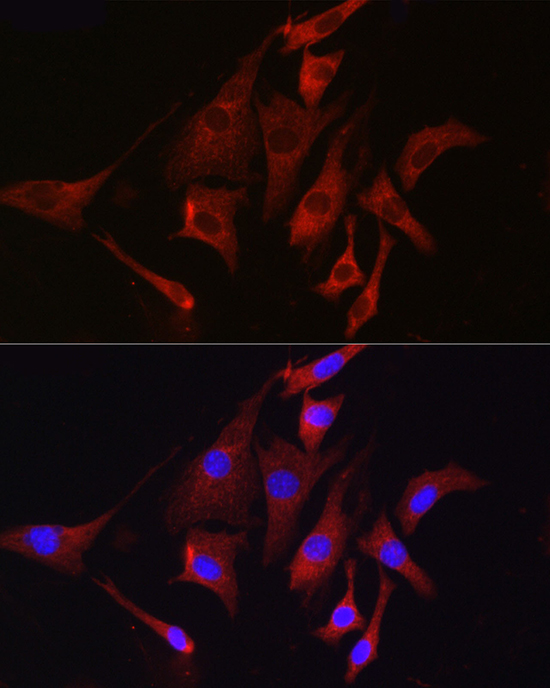
Immunofluorescence analysis of NIH/3T3 cells using TMPRSS2 Rabbit pAb (A1979) at dilution of 1:50 (40x lens). Secondary antibody: Cy3-conjugated Goat anti-Rabbit IgG (H+L) (AS007) at 1:500 dilution. Blue: DAPI for nuclear staining. |
Product Guarantee and Expert Support
