
RPL10A Rabbit mAb, Unconjugated, Monoclonal
Catalog Number:
ABB-A20944
- Images (9)

| Article Name: | RPL10A Rabbit mAb, Unconjugated, Monoclonal |
| Biozol Catalog Number: | ABB-A20944 |
| Supplier Catalog Number: | A20944 |
| Alternative Catalog Number: | ABB-A20944-20UL,ABB-A20944-100UL |
| Manufacturer: | ABclonal |
| Host: | Rabbit |
| Category: | Antikörper |
| Application: | ELISA, IF, IHC-P, WB |
| Species Reactivity: | Human |
| Immunogen: | Recombinant protein (or fragment).This information is considered to be commercially sensitive. |
| Conjugation: | Unconjugated |
| Alternative Names: | uL1, L10A, CSA19, NEDD6, Csa-19, RPL10A |
| Ribosomes, the organelles that catalyze protein synthesis, consist of a small 40S subunit and a large 60S subunit. Together these subunits are composed of 4 RNA species and approximately 80 structurally distinct proteins. This gene encodes a ribosomal protein that is a component of the 60S subunit. The protein belongs to the L1P family of ribosomal proteins. It is located in the cytoplasm. The expression of this gene is downregulated in the thymus by cyclosporin-A (CsA), an immunosuppressive drug. Studies in mice have shown that the expression of the ribosomal protein L10a gene is downregulated in neural precursor cells during development. This gene previously was referred to as NEDD6 (neural precursor cell expressed, developmentally downregulated 6), but it has been renamed RPL10A (ribosomal protein 10a). As is typical for genes encoding ribosomal proteins, there are multiple processed pseudogenes of this gene dispersed through the genome. |
| Application Dilute: | WB,1:500 - 1:1000|IHC-P,1:200 - 1:800|IF/ICC,1:50 - 1:200|ELISA,Recommended starting concentration is 1 µg/mL. Please optimize the concentration based on your specific assay requirements. |
| Application Notes: | Cross-Reactivity: Human,Mouse,Rat. ResearchArea: Epigenetics Nuclear Signaling,RNA Binding. Shipping: Ice Bag |
|
|
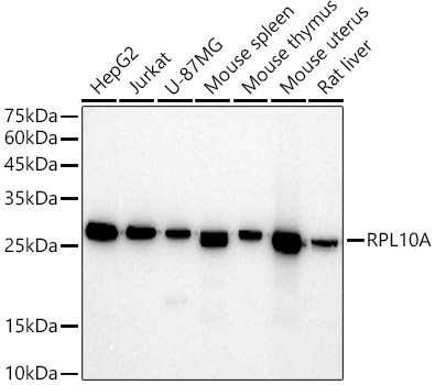
Western blot analysis of various lysates using RPL10A Rabbit mAb (A20944) at 1:500 dilution. Secondary antibody: HRP-conjugated Goat anti-Rabbit IgG (H+L) (AS014) at 1:10000 dilution. Lysates/proteins: 25µg per lane. Blocking buffer: 3% nonfat dry milk in TBST. Detection: ECL Basic Kit (RM00020). Exposure time: 30s. |
|
|
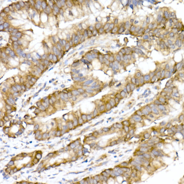
Immunohistochemistry analysis of paraffin-embedded Human lung adenocarcinoma tissue using RPL10A Rabbit mAb (A20944) at a dilution of 1:200 (40x lens). High pressure antigen retrieval performed with 0.01M Citrate Buffer (pH 6.0) prior to IHC staining. |
|
|
Immunohistochemistry analysis of paraffin-embedded Human spleen tissue using RPL10A Rabbit mAb (A20944) at a dilution of 1:200 (40x lens). High pressure antigen retrieval performed with 0.01M Citrate Buffer (pH 6.0) prior to IHC staining. |
|
|
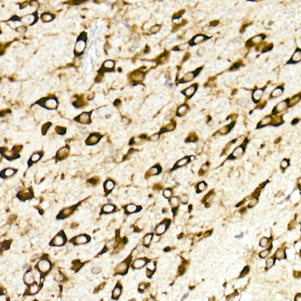
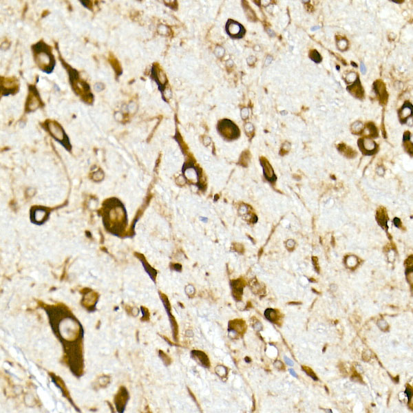
Immunohistochemistry analysis of paraffin-embedded Moue brain tissue using RPL10A Rabbit mAb (A20944) at a dilution of 1:200 (40x lens). High pressure antigen retrieval performed with 0.01M Citrate Buffer (pH 6.0) prior to IHC staining. |
|
|
Immunohistochemistry analysis of paraffin-embedded Moue testis tissue using RPL10A Rabbit mAb (A20944) at a dilution of 1:200 (40x lens). High pressure antigen retrieval performed with 0.01M Citrate Buffer (pH 6.0) prior to IHC staining. |
|
|
Immunohistochemistry analysis of paraffin-embedded Rat brain tissue using RPL10A Rabbit mAb (A20944) at a dilution of 1:200 (40x lens). High pressure antigen retrieval performed with 0.01M Citrate Buffer (pH 6.0) prior to IHC staining. |
|
|
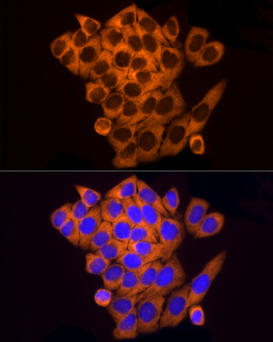
Immunofluorescence analysis of HeLa cells using RPL10A Rabbit mAb (A20944) at dilution of 1:100 (40x lens). Secondary antibody: Cy3-conjugated Goat anti-Rabbit IgG (H+L) (AS007) at 1:500 dilution. Blue: DAPI for nuclear staining. |
|
|
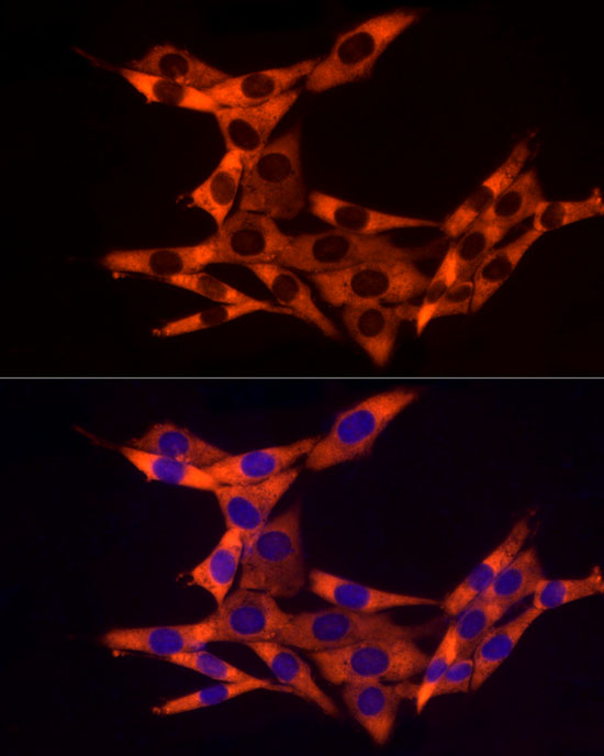
Immunofluorescence analysis of NIH/3T3 cells using RPL10A Rabbit mAb (A20944) at dilution of 1:100 (40x lens). Secondary antibody: Cy3-conjugated Goat anti-Rabbit IgG (H+L) (AS007) at 1:500 dilution. Blue: DAPI for nuclear staining. |
|
|
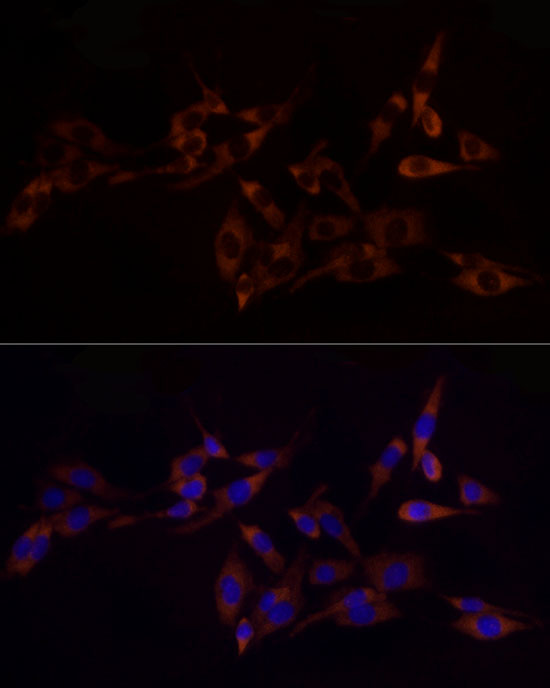
Immunofluorescence analysis of PC-12 cells using RPL10A Rabbit mAb (A20944) at dilution of 1:100 (40x lens). Secondary antibody: Cy3-conjugated Goat anti-Rabbit IgG (H+L) (AS007) at 1:500 dilution. Blue: DAPI for nuclear staining. |
Product Guarantee and Expert Support
