
EIF4G2/p97 Rabbit mAb, Unconjugated, Monoclonal
Catalog Number:
ABB-A21193
- Images (9)

| Article Name: | EIF4G2/p97 Rabbit mAb, Unconjugated, Monoclonal |
| Biozol Catalog Number: | ABB-A21193 |
| Supplier Catalog Number: | A21193 |
| Alternative Catalog Number: | ABB-A21193-100UL,ABB-A21193-20UL |
| Manufacturer: | ABclonal |
| Host: | Rabbit |
| Category: | Antikörper |
| Application: | ELISA, IHC-P, IP, WB |
| Species Reactivity: | Human |
| Immunogen: | Recombinant protein (or fragment).This information is considered to be commercially sensitive. |
| Conjugation: | Unconjugated |
| Alternative Names: | P97, AAG1, DAP5, NAT1, EIF4G2/p97 |
| Translation initiation is mediated by specific recognition of the cap structure by eukaryotic translation initiation factor 4F (eIF4F), which is a cap binding protein complex that consists of three subunits: eIF4A, eIF4E and eIF4G. The protein encoded by this gene shares similarity with the C-terminal region of eIF4G that contains the binding sites for eIF4A and eIF3, eIF4G, in addition, contains a binding site for eIF4E at the N-terminus. Unlike eIF4G, which supports cap-dependent and independent translation, this gene product functions as a general repressor of translation by forming translationally inactive complexes. In vitro and in vivo studies indicate that translation of this mRNA initiates exclusively at a non-AUG (GUG) codon. Alternatively spliced transcript variants encoding different isoforms of this gene have been described. |
| Application Dilute: | WB,1:5000 - 1:20000|IHC-P,1:500 - 1:1000|IP,0.5µg-4µg antibody for 200µg-600µg extracts of whole cells|ELISA,Recommended starting concentration is 1 µg/mL. Please optimize the concentration based on your specific assay requirements. |
| Application Notes: | Cross-Reactivity: Human,Mouse,Rat. ResearchArea: Epigenetics Nuclear Signaling,RNA Binding,Cell Biology Developmental Biology,Apoptosis. Shipping: Ice Bag |
|
|
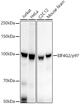
Western blot analysis of various lysates, using EIF4G2/p97 Rabbit mAb (A21193) at 1:5000 dilution. Secondary antibody: HRP-conjugated Goat anti-Rabbit IgG (H+L) (AS014) at 1:10000 dilution. Lysates/proteins: 25µg per lane. Blocking buffer: 3% nonfat dry milk in TBST. Detection: ECL Basic Kit (RM00020). Exposure time: 30s. |
|
|
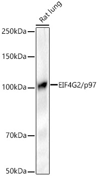
Western blot analysis of lysates from Rat lung, using EIF4G2/p97 Rabbit mAb (A21193) at 1:5000 dilution. Secondary antibody: HRP-conjugated Goat anti-Rabbit IgG (H+L) (AS014) at 1:10000 dilution. Lysates/proteins: 25µg per lane. Blocking buffer: 3% nonfat dry milk in TBST. Detection: ECL Basic Kit (RM00020). Exposure time: 90s. |
|
|
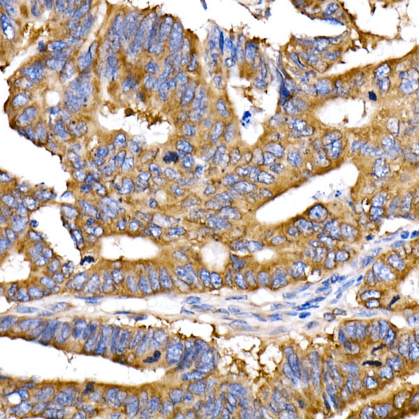
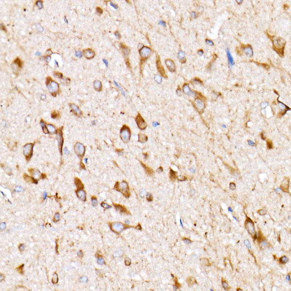
Immunohistochemistry analysis of paraffin-embeddedMouse brain tissue usingEIF4G2/p97 Rabbit mAb(A21193) at a dilution of 1:1000 (40x lens).High pressure antigen retrieval was performed with 0.01 M citrate buffer (pH 6.0) prior to IHC staining. |
|
|
Immunohistochemistry analysis of paraffin-embeddedHuman pancreas tissue usingEIF4G2/p97 Rabbit mAb(A21193) at a dilution of 1:1000 (40x lens).High pressure antigen retrieval was performed with 0.01 M citrate buffer (pH 6.0) prior to IHC staining. |
|
|
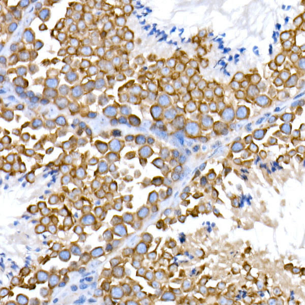
Immunohistochemistry analysis of paraffin-embeddedRat testis tissue usingEIF4G2/p97 Rabbit mAb(A21193) at a dilution of 1:1000 (40x lens).High pressure antigen retrieval was performed with 0.01 M citrate buffer (pH 6.0) prior to IHC staining. |
|
|
Immunohistochemistry analysis of paraffin-embeddedHuman breast cancer tissue usingEIF4G2/p97 Rabbit mAb(A21193) at a dilution of 1:1000 (40x lens).High pressure antigen retrieval was performed with 0.01 M citrate buffer (pH 6.0) prior to IHC staining. |
|
|
Immunohistochemistry analysis of paraffin-embeddedRat brain tissue usingEIF4G2/p97 Rabbit mAb(A21193) at a dilution of 1:1000 (40x lens).High pressure antigen retrieval was performed with 0.01 M citrate buffer (pH 6.0) prior to IHC staining. |
|
|
Immunohistochemistry analysis of paraffin-embeddedMouse testis tissue usingEIF4G2/p97 Rabbit mAb(A21193) at a dilution of 1:1000 (40x lens).High pressure antigen retrieval was performed with 0.01 M citrate buffer (pH 6.0) prior to IHC staining. |
|
|
Immunoprecipitation of EIF4G2/p97 from 300 µg extracts of HeLa cells was performed using 2 µg of EIF4G2/p97 Rabbit mAb (A21193). Rabbit IgG isotype control (AC005) was used to precipitate the Control IgG sample. IP samples were eluted with 1X reducing Laemmli Buffer. The Input lane represents 10% of the total input. Western blot analysis of immunoprecipitates was conducted using EIF4G2/p97 Rabbit mAb (A21193) at a dilution of 1:10000. |
Product Guarantee and Expert Support
