
[KO Validated] NQO1 Rabbit mAb, Unconjugated, Monoclonal
Catalog Number:
ABB-A22290
- Images (9)

| Article Name: | [KO Validated] NQO1 Rabbit mAb, Unconjugated, Monoclonal |
| Biozol Catalog Number: | ABB-A22290 |
| Supplier Catalog Number: | A22290 |
| Alternative Catalog Number: | ABB-A22290-100UL,ABB-A22290-20UL,ABB-A22290-500UL,ABB-A22290-1000UL |
| Manufacturer: | ABclonal |
| Host: | Rabbit |
| Category: | Antikörper |
| Application: | ELISA, IF, IHC-P, WB |
| Species Reactivity: | Human |
| Immunogen: | Recombinant protein (or fragment).This information is considered to be commercially sensitive. |
| Conjugation: | Unconjugated |
| Alternative Names: | DTD, QR1, DHQU, DIA4, NMOR1, NMORI, [KD Validated] NQO1 |
| This gene is a member of the NAD(P)H dehydrogenase (quinone) family and encodes a cytoplasmic 2-electron reductase. This FAD-binding protein forms homodimers and reduces quinones to hydroquinones. This proteins enzymatic activity prevents the one electron reduction of quinones that results in the production of radical species. Mutations in this gene have been associated with tardive dyskinesia (TD), an increased risk of hematotoxicity after exposure to benzene, and susceptibility to various forms of cancer. Altered expression of this protein has been seen in many tumors and is also associated with Alzheimers disease (AD). Alternate transcriptional splice variants, encoding different isoforms, have been characterized. |
| Clonality: | Monoclonal |
| Clone Designation: | [ARC56754] |
| Molecular Weight: | 31 kDa |
| NCBI: | 1728 |
| UniProt: | P15559 |
| Purity: | Affinity purification |
| Sequence: | MVGRRALIVLAHSERTSFNYAMKEAAAAALKKKGWEVVESDLYAMNFNPIISRKDITGKLKDPANFQYPAESVLAYKEGHLSPDIVAEQKKLEAADLVIFQFPLQWFGVPAILKGWFERVFIGEFAYTYAAMYDKGPFRSKKAVLSITTGGSGSMYSLQGIHGDMNVILWPIQSGILHFCGFQVLEPQLTYSIGHTPADARIQILEGWKKRLENIWDETPLYFAPSSLFDLNFQAGFLMKKEVQDEEKNKKFGLS |
| Target: | NQO1 |
| Antibody Type: | Primary Antibody |
| Application Dilute: | WB,1:20000 - 1:200000|IF-P,1:100 - 1:500|IHC-P,1:500 - 1:2000|ELISA,Recommended starting concentration is 1 µg/mL. Please optimize the concentration based on your specific assay requirements. |
| Application Notes: | Cross-Reactivity: Human,Mouse,Rat. ResearchArea: Epigenetics Nuclear Signaling,RNA Binding,Cancer,Signal Transduction,Endocrine Metabolism,Drug metabolism. Shipping: Ice Bag |
|
|
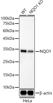
Western blot analysis of lysates from wild type (WT) and NQO1 knockout (KO) HeLa cells using [KO Validated] NQO1 Rabbit mAb (A22290) at 1:120000 dilution incubated overnight at 4°C. Secondary antibody: HRP-conjugated Goat anti-Rabbit IgG (H+L) (AS014) at 1:10000 dilution. Lysates/proteins: 25 µg per lane. Blocking buffer: 3% nonfat dry milk in TBST. Detection: ECL Basic Kit (RM00020). Exposure time: 20s. |
|
|
Immunohistochemistry analysis of paraffin-embedded Human breast cancer tissue using [KO Validated] NQO1 Rabbit mAb (A22290) at a dilution of 1:1000 (40x lens). High pressure antigen retrieval performed with 0.01M Tris-EDTA Buffer (pH 9.0) prior to IHC staining. |
|
|
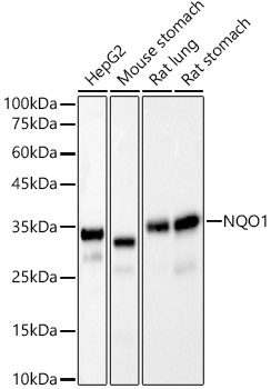
Western blot analysis of various lysates, using [KO Validated] NQO1 Rabbit mAb (A22290) at1:20000 dilution. Secondary antibody: HRP-conjugated Goat anti-Rabbit IgG (H+L) (AS014) at 1:10000 dilution. Lysates/proteins: 25µg per lane. Blocking buffer: 3% nonfat dry milk in TBST. Detection: ECL Basic Kit (RM00020). Exposure time: 10s. |
|
|
Immunohistochemistry analysis of paraffin-embedded Human colon carcinoma tissue using [KO Validated] NQO1 Rabbit mAb (A22290) at a dilution of 1:1000 (40x lens). High pressure antigen retrieval performed with 0.01M Tris-EDTA Buffer (pH 9.0) prior to IHC staining. |
|
|
Immunohistochemistry analysis of paraffin-embedded Human stomach tissue using [KO Validated] NQO1 Rabbit mAb (A22290) at a dilution of 1:1000 (40x lens). High pressure antigen retrieval performed with 0.01M Tris-EDTA Buffer (pH 9.0) prior to IHC staining. |
|
|
Immunohistochemistry analysis of paraffin-embedded Human testis tissue using [KO Validated] NQO1 Rabbit mAb (A22290) at a dilution of 1:1000 (40x lens). High pressure antigen retrieval performed with 0.01M Tris-EDTA Buffer (pH 9.0) prior to IHC staining. |
|
|
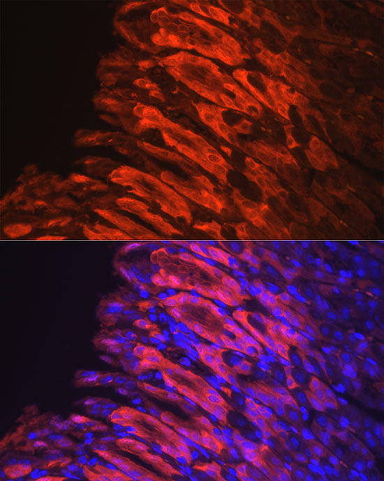
Confocal imaging of paraffin-embedded human stomach using [KO Validated] NQO1 Rabbit mAb (A22290, dilution 1:200) followed by a further incubation with Cy3 Goat Anti-Rabbit IgG (H+L) (AS007, dilution 1:500) (Red). DAPI was used for nuclear staining (Blue). Objective: 40x.Perform high pressure antigen retrieval with 0.01M citrate buffer (pH 6.0) prior to IF staining. |
|
|
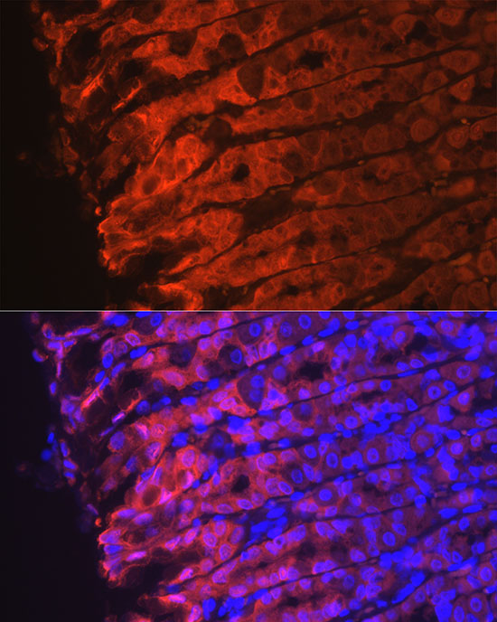
Confocal imaging of paraffin-embedded mouse stomach using [KO Validated] NQO1 Rabbit mAb (A22290, dilution 1:200) followed by a further incubation with Cy3 Goat Anti-Rabbit IgG (H+L) (AS007, dilution 1:500) (Red). DAPI was used for nuclear staining (Blue). Objective: 40x.Perform high pressure antigen retrieval with 0.01M citrate buffer (pH 6.0) prior to IF staining. |
|
|
Confocal imaging of paraffin-embedded rat stomach using [KO Validated] NQO1 Rabbit mAb (A22290, dilution 1:200) followed by a further incubation with Cy3 Goat Anti-Rabbit IgG (H+L) (AS007, dilution 1:500) (Red). DAPI was used for nuclear staining (Blue). Objective: 40x.Perform high pressure antigen retrieval with 0.01M citrate buffer (pH 6.0) prior to IF staining. |
Product Guarantee and Expert Support
