
[KO Validated] NF-kB p65/RelA Rabbit mAb, Unconjugated, Monoclonal
Catalog Number:
ABB-A22331
- Images (9)


| Article Name: | [KO Validated] NF-kB p65/RelA Rabbit mAb, Unconjugated, Monoclonal |
| Biozol Catalog Number: | ABB-A22331 |
| Supplier Catalog Number: | A22331 |
| Alternative Catalog Number: | ABB-A22331-100UL,ABB-A22331-20UL,ABB-A22331-500UL,ABB-A22331-1000UL |
| Manufacturer: | ABclonal |
| Host: | Rabbit |
| Category: | Antikörper |
| Application: | ChIP, ELISA, IF, IP, WB |
| Species Reactivity: | Human |
| Immunogen: | Synthetic peptide. This information is considered to be commercially sensitive. |
| Conjugation: | Unconjugated |
| Alternative Names: | p65, CMCU, NFKB3, AIF3BL3, lA |
| NF-kappa-B is a ubiquitous transcription factor involved in several biological processes. It is held in the cytoplasm in an inactive state by specific inhibitors. Upon degradation of the inhibitor, NF-kappa-B moves to the nucleus and activates transcription of specific genes. NF-kappa-B is composed of NFKB1 or NFKB2 bound to either REL, RELA, or RELB. The most abundant form of NF-kappa-B is NFKB1 complexed with the product of this gene, RELA. Four transcript variants encoding different isoforms have been found for this gene. |
| Clonality: | Monoclonal |
| Clone Designation: | [ARC51088] |
| Molecular Weight: | 58kDa/59kDa/60kDa |
| NCBI: | 5970 |
| UniProt: | Q04206 |
| Purity: | Affinity purification |
| Sequence: | LGALLGNSTDPAVFTDLASVDNSEFQQLLNQGIPVAPHTTEPMLMEYPEAITRLVTGAQRPPDPAPAPLGAPGLPNGLLSGDEDFSSIADMDFSALLSQISS |
| Target: | RELA |
| Antibody Type: | Primary Antibody |
| Application Dilute: | WB,1:5000 - 1:20000|IF/ICC,1:500 - 1:2000|IP,0.5µg-4µg antibody for 200µg-500µg extracts of whole cells|ChIP,5µg antibody for 10µg-15µg of Chromatin|ELISA,Recommended starting concentration is 1 µg/mL. Please optimize the concentration based on your speci |
| Application Notes: | Cross-Reactivity: Human,Mouse,Rat,Monkey. ResearchArea: Epigenetics Nuclear Signaling,Transcription Factors,Protein phosphorylation,Cancer,Signal Transduction,Cell Biology Developmental Biology,Apoptosis,Inhibition of Apoptosis,Death Receptor Signaling Pathway,Endocrine Metabolism,Endocrine and metabolic diseases,Obesity,Immunology Inflammation,B Cell Receptor Signaling Pathway,T Cell Receptor Signaling Pathway,Jak-Stat-IL-6 Receptor Signaling Pathway,NF-kB Signaling Pathway,Toll-like Receptor Signaling Pathway,Neuroscience,Neurodegenerative Diseases,Amyloid Plaque and Neurofibrillary Tangle Formation in Alzheimers Disease,Cardiovascular. Shipping: Ice Bag |
|
|
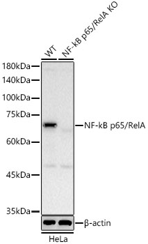
Western blot analysis of lysates from wild type(WT) and NF-kB p65/RelA knockout (KO) HeLa cells, using [KO Validated] NF-kB p65/RelA Rabbit mAb (A22331) at 1:10000 dilution. Secondary antibody: HRP-conjugated Goat anti-Rabbit IgG (H+L) (AS014) at 1:10000 dilution. Lysates/proteins: 25µg per lane. Blocking buffer: 3% nonfat dry milk in TBST. Detection: ECL Basic Kit (RM00020). Exposure time: 10s. |
|
|
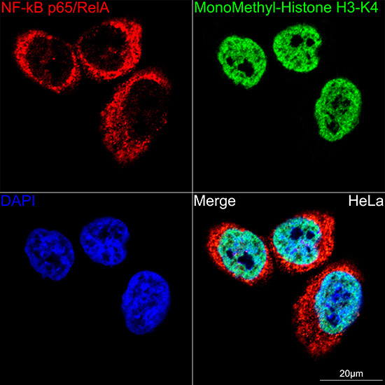
Confocal imaging of PC-12 cells (treated with TNF-alpha) and PC-12 cells (untreated) cells using [KO Validated] NF-kB p65/RelA Rabbit mAb (A22331, dilution 1:2000) followed by a further incubation with Cy3 Goat Anti-Rabbit IgG (H+L) (AS007, dilution 1:500) (Red). The cells were counterstained with alpha-Tubulin Mouse mAb (AC012, dilution 1:400) followed by incubation with ABflo 488-conjugated Goat Anti-Mouse IgG (H+L) Ab (AS076, dilution 1:500) (Green). DAPI was used for nuclear staining (Blue). Objective: 100x. |
|
|
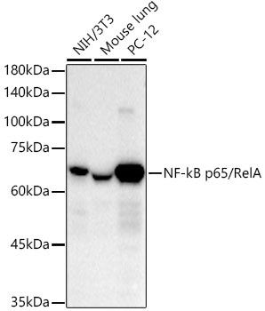
Western blot analysis of various lysates using [KO Validated] NF-kB p65/RelA Rabbit mAb (A22331) at 1:10000 dilution. Secondary antibody: HRP-conjugated Goat anti-Rabbit IgG (H+L) (AS014) at 1:10000 dilution. Lysates/proteins: 25µg per lane. Blocking buffer: 3% nonfat dry milk in TBST. Detection: ECL Basic Kit (RM00020). Exposure time: 30s. |
|
|
Confocal imaging of HT-1080 cells (treated with TNF-alpha) and HT-1080 cells (untreated) cells using [KO Validated] NF-kB p65/RelA Rabbit mAb (A22331, dilution 1:2000) followed by a further incubation with Cy3 Goat Anti-Rabbit IgG (H+L) (AS007, dilution 1:500) (Red). The cells were counterstained with alpha-Tubulin Mouse mAb (AC012, dilution 1:400) followed by incubation with ABflo 488-conjugated Goat Anti-Mouse IgG (H+L) Ab (AS076, dilution 1:500) (Green). DAPI was used for nuclear staining (Blue). Objective: 100x. |
|
|
Confocal imaging of NIH/3T3 cells (treated with TNF-alpha) and NIH/3T3 cells (untreated) cells using [KO Validated] NF-kB p65/RelA Rabbit mAb (A22331, dilution 1:2000) followed by a further incubation with Cy3 Goat Anti-Rabbit IgG (H+L) (AS007, dilution 1:500) (Red). The cells were counterstained with alpha-Tubulin Mouse mAb (AC012, dilution 1:400) followed by incubation with ABflo 488-conjugated Goat Anti-Mouse IgG (H+L) Ab (AS076, dilution 1:500) (Green). DAPI was used for nuclear staining (Blue). Objective: 100x. |
|
|
Immunoprecipitation of [KO Validated] NF-kB p65/RelA Rabbit mAb from 500 µg extracts of HeLa cells was performed using 2 µg of [KO Validated] NF-kB p65/RelA Rabbit mAb (A22331). Rabbit IgG isotype control (AC042) was used to precipitate the Control IgG sample. IP samples were eluted with 1X Laemmli Buffer. The Input lane represents 10% of the total input. Western blot analysis of immunoprecipitates was conducted using [KO Validated] NF-kB p65/RelA Rabbit mAb (A22331) at a dilution of 1:10000. |
|
|
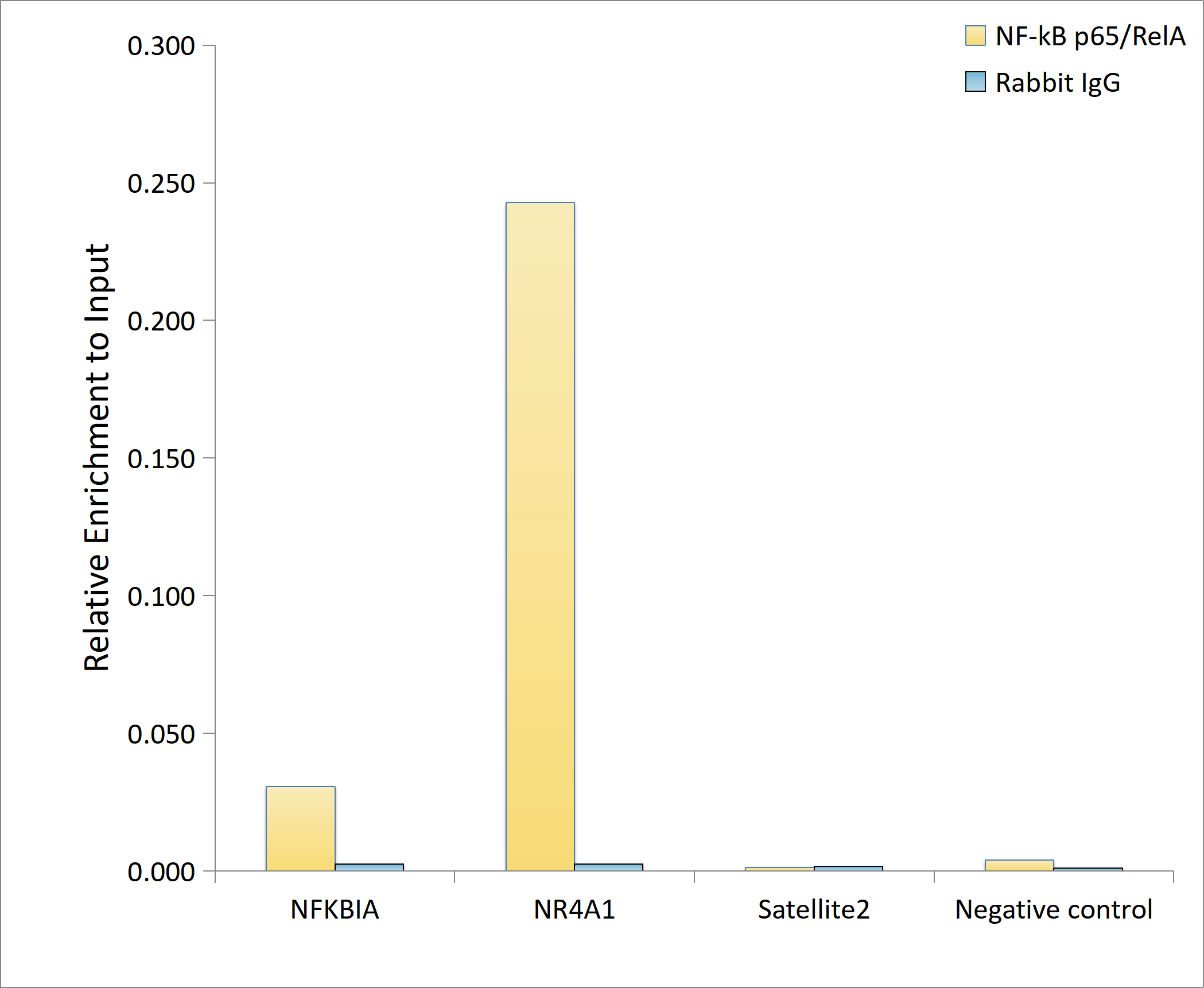
Chromatin immunoprecipitation analysis of extracts of HT-1080 cells, HT-1080 cells were treated by TNF-alpha (20 ng/ml) at 37°C for 30 minutes, using [KO Validated] NF-kB p65/RelA Rabbit mAb (A22331) and rabbit IgG.The amount of immunoprecipitated DNA was checked by quantitative PCR. Histogram was constructed by the ratios of the immunoprecipitated DNA to the input. |
|
|
Immunoprecipitation of [KO Validated] NF-kB p65/RelA Rabbit mAb from 500 µg extracts of HeLa cells was performed using 2 µg of [KO Validated] NF-kB p65/RelA Rabbit mAb (A22331). Rabbit IgG isotype control (AC042) was used to precipitate the Control IgG sample. IP samples were eluted with 1X Laemmli Buffer. The Input lane represents 10% of the total input. Western blot analysis of immunoprecipitates was conducted using [KO Validated] NF-kB p65/RelA Rabbit mAb (A22331) at a dilution of 1:10000. |
|
|
Chromatin immunoprecipitation analysis of extracts of HT-1080 cells, HT-1080 cells were treated by TNF-alpha (20 ng/ml) at 37°C for 30 minutes, using [KO Validated] NF-kB p65/RelA Rabbit mAb (A22331) and rabbit IgG.The amount of immunoprecipitated DNA was checked by quantitative PCR. Histogram was constructed by the ratios of the immunoprecipitated DNA to the input. |
Product Guarantee and Expert Support
