
ACVR1B Rabbit mAb, Unconjugated, Monoclonal
Catalog Number:
ABB-A2279
- Images (9)

| Article Name: | ACVR1B Rabbit mAb, Unconjugated, Monoclonal |
| Biozol Catalog Number: | ABB-A2279 |
| Supplier Catalog Number: | A2279 |
| Alternative Catalog Number: | ABB-A2279-20UL,ABB-A2279-500UL,ABB-A2279-1000UL,ABB-A2279-100UL |
| Manufacturer: | ABclonal |
| Host: | Rabbit |
| Category: | Antikörper |
| Application: | ELISA, IF, IHC-P, WB |
| Species Reactivity: | Human |
| Immunogen: | Synthetic peptide. This information is considered to be commercially sensitive. |
| Conjugation: | Unconjugated |
| Alternative Names: | ALK4, SKR2, ACTRIB, ACVRLK4, ACVR1B |
| This gene encodes an activin A type IB receptor. Activins are dimeric growth and differentiation factors which belong to the transforming growth factor-beta (TGF-beta) superfamily of structurally related signaling proteins. Activins signal through a heteromeric complex of receptor serine kinases which include at least two type I and two type II receptors. This protein is a type I receptor which is essential for signaling. Mutations in this gene are associated with pituitary tumors. Alternate splicing results in multiple transcript variants. |
| Application Dilute: | WB,1:500 - 1:2000|IHC-P,1:50 - 1:200|IF/ICC,1:50 - 1:200|ELISA,Recommended starting concentration is 1 µg/mL. Please optimize the concentration based on your specific assay requirements. |
| Application Notes: | Cross-Reactivity: Human,Mouse,Rat. ResearchArea: Signal Transduction,Cell Biology Developmental Biology,Apoptosis,Cytoskeleton,Microfilaments,Stem Cells. Shipping: Ice Bag |
|
|
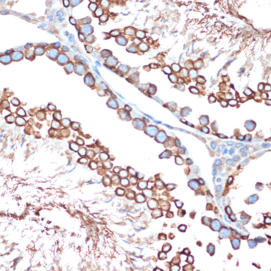
Immunohistochemistry analysis of paraffin-embedded Rat testis using ACVR1B Rabbit mAb (A2279) at dilution of 1:100 (40x lens). Microwave antigen retrieval performed with 0.01M Tris/EDTA Buffer (pH 9.0) prior to IHC staining. |
|
|
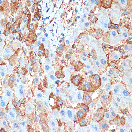
Immunohistochemistry analysis of paraffin-embedded Human liver using ACVR1B Rabbit mAb (A2279) at dilution of 1:100 (40x lens). Microwave antigen retrieval performed with 0.01M Tris/EDTA Buffer (pH 9.0) prior to IHC staining. |
|
|
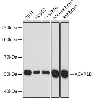
Western blot analysis of various lysates using ACVR1B Rabbit mAb (A2279) at 1:1000 dilution. Secondary antibody: HRP-conjugated Goat anti-Rabbit IgG (H+L) (AS014) at 1:10000 dilution. Lysates/proteins: 25µg per lane. Blocking buffer: 3% nonfat dry milk in TBST. Detection: ECL Basic Kit (RM00020). Exposure time: 3min. |
|
|
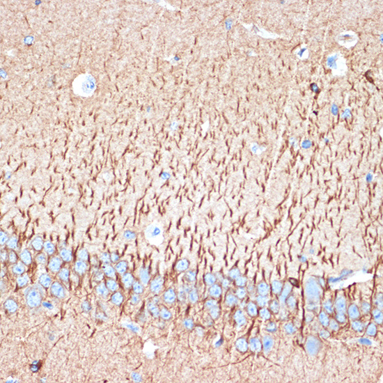
Immunohistochemistry analysis of paraffin-embedded Mouse brain using ACVR1B Rabbit mAb (A2279) at dilution of 1:100 (40x lens). Microwave antigen retrieval performed with 0.01M Tris/EDTA Buffer (pH 9.0) prior to IHC staining. |
|
|
Immunohistochemistry analysis of paraffin-embedded Human cervix cancer tissue using ACVR1B Rabbit mAb (A22347) at a dilution of 1:200 (40x lens). High pressure antigen retrieval performed with 0.01M Citrate buffer (pH 6.0) prior to IHC staining. |
|
|
Immunohistochemistry analysis of paraffin-embedded Human colon tissue using ACVR1B Rabbit mAb (A22347) at a dilution of 1:200 (40x lens). High pressure antigen retrieval performed with 0.01M Citrate buffer (pH 6.0) prior to IHC staining. |
|
|
Immunohistochemistry analysis of paraffin-embedded Mouse kidney tissue using ACVR1B Rabbit mAb (A22347) at a dilution of 1:200 (40x lens). High pressure antigen retrieval performed with 0.01M Citrate buffer (pH 6.0) prior to IHC staining. |
|
|
Immunohistochemistry analysis of paraffin-embedded Mouse testis tissue using ACVR1B Rabbit mAb (A22347) at a dilution of 1:200 (40x lens). High pressure antigen retrieval performed with 0.01M Citrate buffer (pH 6.0) prior to IHC staining. |
|
|
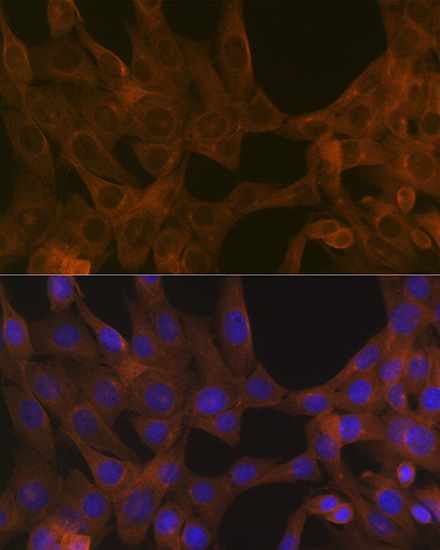
Confocal imaging of NIH/3T3 cells usingACVR1B Rabbit mAb (A2279,dilution 1:100) followed by a further incubation with Cy3 Goat Anti-Rabbit IgG (H+L) (AS007,dilution 1:500)(Red).The cells were counterstained with alpha-Tubulin Mouse mAb (AC012, dilution 1:400) followed by incubation with ABflo 488-conjugated Goat Anti-Mouse IgG (H+L) Ab (AS076, dilution 1:500) (Green).DAPI was used for nuclear staining (Blue). Objective: 100x. |
Product Guarantee and Expert Support
