
CD45 Rabbit mAb, Unconjugated, Monoclonal
Catalog Number:
ABB-A23503
- Images (9)

| Article Name: | CD45 Rabbit mAb, Unconjugated, Monoclonal |
| Biozol Catalog Number: | ABB-A23503 |
| Supplier Catalog Number: | A23503 |
| Alternative Catalog Number: | ABB-A23503-500UL,ABB-A23503-100UL,ABB-A23503-1000UL,ABB-A23503-20UL |
| Manufacturer: | ABclonal |
| Host: | Rabbit |
| Category: | Antikörper |
| Application: | ELISA, IHC, IHC-P, WB |
| Species Reactivity: | Human |
| Immunogen: | Recombinant protein (or fragment).This information is considered to be commercially sensitive. |
| Conjugation: | Unconjugated |
| Alternative Names: | LCA, LY5, B220, CD45, L-CA, T200, CD45R, GP180, IMD105 |
| The protein encoded by this gene is a member of the protein tyrosine phosphatase (PTP) family. PTPs are known to be signaling molecules that regulate a variety of cellular processes including cell growth, differentiation, mitosis, and oncogenic transformation. This PTP contains an extracellular domain, a single transmembrane segment and two tandem intracytoplasmic catalytic domains, and thus is classified as a receptor type PTP. This PTP has been shown to be an essential regulator of T- and B-cell antigen receptor signaling. It functions through either direct interaction with components of the antigen receptor complexes, or by activating various Src family kinases required for the antigen receptor signaling. This PTP also suppresses JAK kinases, and thus functions as a regulator of cytokine receptor signaling. Alternatively spliced transcripts variants of this gene, which encode distinct isoforms, have been reported. |
| Clonality: | Monoclonal |
| Clone Designation: | [ARC52280] |
| Molecular Weight: | 147kDa |
| NCBI: | 5788 |
| UniProt: | P08575 |
| Purity: | Affinity purification |
| Sequence: | SDAYLNASETTTLSPSGSAVISTTTIATTPSKPTCDEKYANITVDYLYNKETKLFTAKLNVNENVECGNNTCTNNEVHNLTECKNASVSISHNSCTAPDKTLILDVPPGVEKFQLHDCTQVEKADTTICLKWKNIETFTCDTQNITYRFQCGNMIFDNKEIKLENLEPEHEYKCDSEILYNNHKFTNASKIIKTDFG |
| Target: | PTPRC |
| Antibody Type: | Primary Antibody |
| Application Dilute: | WB,1:100 - 1:500|IHC-P,1:5000 - 1:20000|mIHC,1:5000 - 1:20000|ELISA,Recommended starting concentration is 1 µg/mL. Please optimize the concentration based on your specific assay requirements. |
| Application Notes: | Cross-Reactivity: Human. ResearchArea: Signal Transduction,Kinase,Tyrosine kinases,Cell Biology Developmental Biology,Apoptosis,Immunology Inflammation,CDs,B Cell Receptor Signaling Pathway,T Cell Receptor Signaling Pathway,Neuroscience, Cell Type Marker,Stem Cells,Hematopoietic Progenitors. Shipping: Ice Bag |
|
|
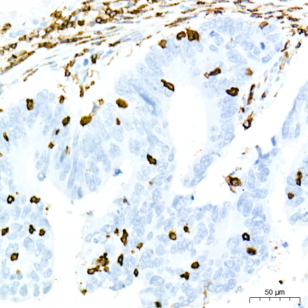
Immunohistochemistry analysis of paraffin-embedded Human colon tissue using CD45 Rabbit mAb (A23503) at a dilution of 1:5000 (40x lens). High pressure antigen retrieval performed with 0.01M Tris-EDTA Buffer (pH 9.0) prior to IHC staining. |
|
|
Immunohistochemistry analysis of paraffin-embedded Human esophagus tissue using CD45 Rabbit mAb (A23503) at a dilution of 1:5000 (40x lens). High pressure antigen retrieval performed with 0.01M Tris-EDTA Buffer (pH 9.0) prior to IHC staining. |
|
|
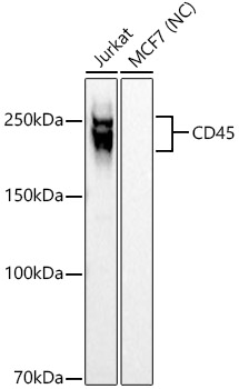
Western blot analysis of various lysates, using CD45 Rabbit mAb (A23503) at 1:500 dilution. Secondary antibody: HRP-conjugated Goat anti-Rabbit IgG (H+L) (AS014) at 1:10000 dilution. Lysates/proteins: 25µg per lane. Blocking buffer: 3% nonfat dry milk in TBST. Detection: ECL Basic Kit (RM00020). Negative control (NC):MCF7 Exposure time: 1s. |
|
|
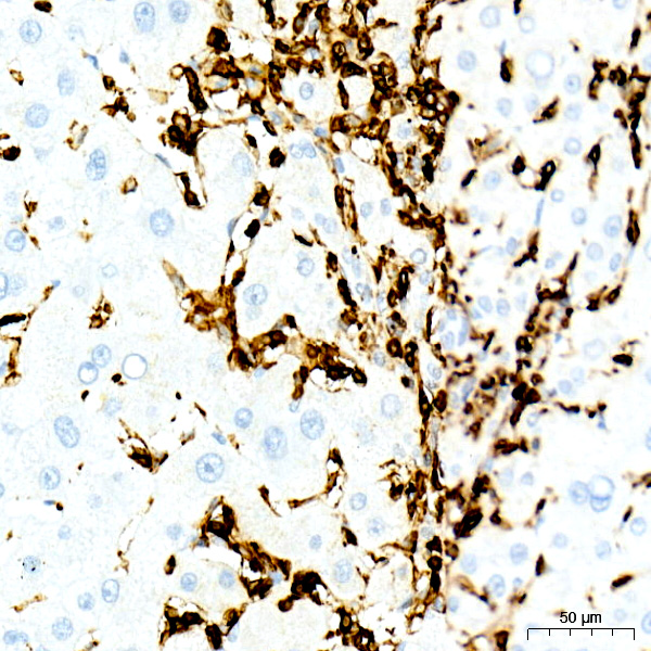
Immunohistochemistry analysis of paraffin-embedded Human liver tissue using CD45 Rabbit mAb (A23503) at a dilution of 1:5000 (40x lens). High pressure antigen retrieval performed with 0.01M Tris-EDTA Buffer (pH 9.0) prior to IHC staining. |
|
|
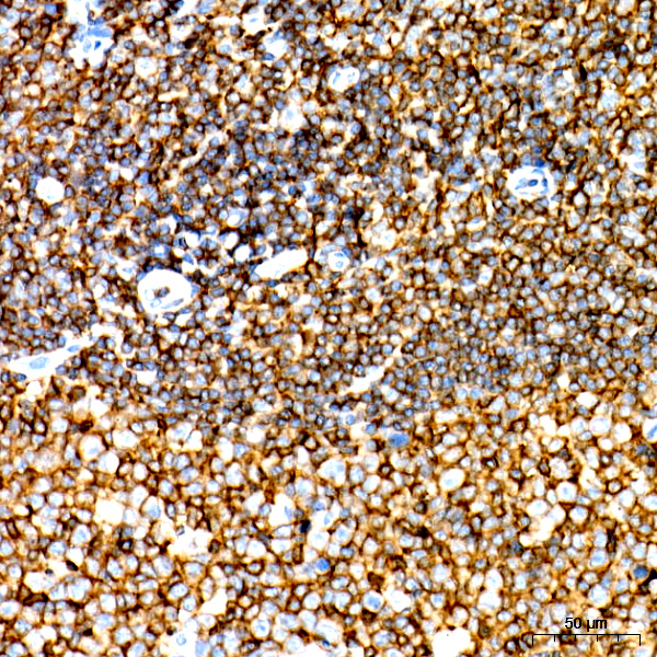
Immunohistochemistry analysis of paraffin-embedded Human tonsil tissue using CD45 Rabbit mAb (A23503) at a dilution of 1:5000 (40x lens). High pressure antigen retrieval performed with 0.01M Tris-EDTA Buffer (pH 9.0) prior to IHC staining. |
|
|
Immunohistochemistry analysis of paraffin-embedded Human spleen tissue using CD45 Rabbit mAb (A23503) at a dilution of 1:5000 (40x lens). High pressure antigen retrieval performed with 0.01M Tris-EDTA Buffer (pH 9.0) prior to IHC staining. |
|
|
The multiplex IHC analysis on paraffin-embedded Human breast cancer tissue using the following specific primary antibodies and tyramide signal amplification (TSA) reagents (RK05903) : CD45 Rabbit mAb (A23503, 1:5000) with TSA-TYR-520 (Green), CD8A Rabbit mAb (A23305PM, 1:1000) with TSA-TYR-570 (Red), and CD3E Rabbit mAb (A24060, 1:200) with TSA-TYR-690 (Magenta). DAPI (Blue) was used for nuclear staining. Prior to multiplex IHC staining, high-pressure antigen retrieval was performed using 0.01M citrate buffer at pH 6.0. The analysis was completed using a 20x objective lens. |
|
|
The multiplex IHC analysis on paraffin-embedded Human colon tissue using the following specific primary antibodies and tyramide signal amplification (TSA) reagents (RK05903) : CD45 Rabbit mAb (A23503, 1:5000) with TSA-TYR-520 (Green), CD8A Rabbit mAb (A23305PM, 1:1000) with TSA-TYR-570 (Red), and CD3E Rabbit mAb (A24060, 1:200) with TSA-TYR-690 (Magenta). DAPI (Blue) was used for nuclear staining. Prior to multiplex IHC staining, high-pressure antigen retrieval was performed using 0.01M citrate buffer at pH 6.0. The analysis was completed using a 20x objective lens. |
|
|
The multiplex IHC analysis on paraffin-embedded Human tonsil tissue using the following specific primary antibodies and tyramide signal amplification (TSA) reagents (RK05903) : CD45 Rabbit mAb (A23503, 1:5000) with TSA-TYR-520 (Green), CD8A Rabbit mAb (A23305PM, 1:1000) with TSA-TYR-570 (Red), and CD3E Rabbit mAb (A24060, 1:200) with TSA-TYR-690 (Magenta). DAPI (Blue) was used for nuclear staining. Prior to multiplex IHC staining, high-pressure antigen retrieval was performed using 0.01M citrate buffer at pH 6.0. The analysis was completed using a 20x objective lens. |
Product Guarantee and Expert Support
