
HMGCS2 Rabbit mAb, Unconjugated, Monoclonal
Catalog Number:
ABB-A23545
- Images (9)

| Article Name: | HMGCS2 Rabbit mAb, Unconjugated, Monoclonal |
| Biozol Catalog Number: | ABB-A23545 |
| Supplier Catalog Number: | A23545 |
| Alternative Catalog Number: | ABB-A23545-100UL,ABB-A23545-20UL |
| Manufacturer: | ABclonal |
| Host: | Rabbit |
| Category: | Antikörper |
| Application: | ELISA, IF, IHC-P, WB |
| Species Reactivity: | Human |
| Immunogen: | Synthetic peptide. This information is considered to be commercially sensitive. |
| Conjugation: | Unconjugated |
| Alternative Names: | HMGCS2, Hydroxymethylglutaryl-CoA synthase, mitochondrial, 3-hydroxy-3-methylglutaryl coenzyme A synthase, 3-hydroxy-3-methylglutaryl-CoA synthase 2 |
| The protein encoded by this gene belongs to the HMG-CoA synthase family. It is a mitochondrial enzyme that catalyzes the first reaction of ketogenesis, a metabolic pathway that provides lipid-derived energy for various organs during times of carbohydrate deprivation, such as fasting. Mutations in this gene are associated with HMG-CoA synthase deficiency. Alternatively spliced transcript variants encoding different isoforms have been found for this gene. |
| Application Dilute: | WB,1:2000 - 1:8000|IF/ICC,1:100 - 1:400|IF-P,1:100 - 1:400|IHC-P,1:1000 - 1:4000|ELISA,Recommended starting concentration is 1 µg/mL. Please optimize the concentration based on your specific assay requirements. |
| Application Notes: | Cross-Reactivity: Human,Mouse,Rat. ResearchArea: Cancer,Signal Transduction,Endocrine Metabolism,Mitochondrial metabolism,Mitochondrial markers,Lipid Metabolism,Cholesterol Metabolism,Cardiovascular,Lipids. Shipping: Ice Bag |
|
|
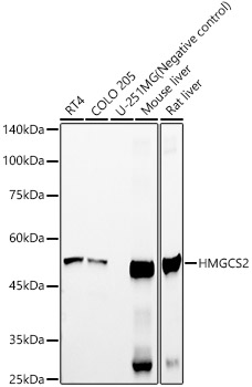
Western blot analysis of various lysates using HMGCS2 Rabbit mAb (A23545) at 1:2000 dilutionincubated overnight at 4°C. Secondary antibody: HRP-conjugated Goat anti-Rabbit IgG (H+L) (AS014) at 1:10000 dilution. Lysates/proteins: 25 µg per lane. Blocking buffer: 3% nonfat dry milk in TBST. Detection: ECL Basic Kit (RM00020). Negative control (NC): U-251 MG Exposure time: 10s. |
|
|
Immunohistochemistry analysis of paraffin-embedded Rat liver tissue using HMGCS2 Rabbit mAb (A23545) at a dilution of 1:2000 (40x lens). High pressure antigen retrieval performed with 0.01M Tris-EDTA Buffer (pH 9.0) prior to IHC staining. |
|
|
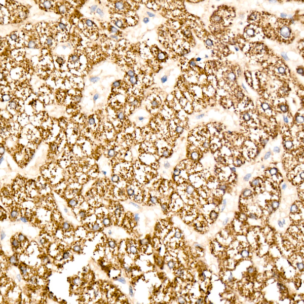
Immunohistochemistry analysis of paraffin-embedded Human liver tissue using HMGCS2 Rabbit mAb (A23545) at a dilution of 1:2000 (40x lens). High pressure antigen retrieval performed with 0.01M Tris-EDTA Buffer (pH 9.0) prior to IHC staining. |
|
|
Immunohistochemistry analysis of paraffin-embedded Mouse liver tissue using HMGCS2 Rabbit mAb (A23545) at a dilution of 1:2000 (40x lens). High pressure antigen retrieval performed with 0.01M Tris-EDTA Buffer (pH 9.0) prior to IHC staining. |
|
|
Immunohistochemistry analysis of paraffin-embedded Human liver cancer tissue using HMGCS2 Rabbit mAb (A23545) at a dilution of 1:2000 (40x lens). High pressure antigen retrieval performed with 0.01M Tris-EDTA Buffer (pH 9.0) prior to IHC staining. |
|
|
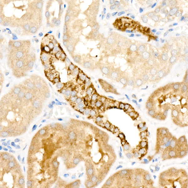
Immunohistochemistry analysis of paraffin-embedded Human kidney tissue using HMGCS2 Rabbit mAb (A23545) at a dilution of 1:2000 (40x lens). High pressure antigen retrieval performed with 0.01M Tris-EDTA Buffer (pH 9.0) prior to IHC staining. |
|
|
Confocal imaging of COLO 205 cells using HMGCS2 Rabbit mAb (A23545, dilution 1:200) followed by a further incubation with Cy3 Goat Anti-Rabbit IgG (H+L) (AS007, dilution 1:500) (Red). DAPI was used for nuclear staining (Blue). Objective: 100x. |
|
|
Confocal imaging of RT-4 cells using HMGCS2 Rabbit mAb (A23545, dilution 1:200) followed by a further incubation with Cy3 Goat Anti-Rabbit IgG (H+L) (AS007, dilution 1:500) (Red). DAPI was used for nuclear staining (Blue). Objective: 100x. |
|
|
Confocal imaging of paraffin-embedded Human liver tissue using HMGCS2 Rabbit mAb (A23545, dilution 1:200) followed by a further incubation with Cy3 Goat Anti-Rabbit IgG (H+L) (AS007, dilution 1:500) (Red). DAPI was used for nuclear staining (Blue). High pressure antigen retrieval performed with 0.01M Citrate Buffer (pH 6.0) prior to IF staining. Objective: 40x. |
Product Guarantee and Expert Support
