Predicted to enable G protein-coupled receptor activity. Predicted to be involved in adenylate cyclase-activating G protein-coupled receptor signaling pathway. Predicted to act upstream of or within G protein-coupled receptor signaling pathway and adaptive immune response. Located in external side of plasma membrane. Is expressed in several structures, including cardiovascular system, central nervous system, genitourinary system, hemolymphoid system, and intestine. Orthologous to human ADGRE1 (adhesion G protein-coupled receptor E1).
IF-P,1:100 - 1:400|IHC-P,1:300 - 1:1200|ELISA,Recommended starting concentration is 1 µg/mL. Please optimize the concentration based on your specific assay requirements.
Application Notes:
Cross-Reactivity: Mouse,Rat. ResearchArea: Immunology,Innate Immunity,Macrophage/Inflamm. Shipping: Ice Bag
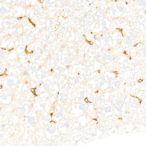
Immunohistochemistry analysis of paraffin-embedded Mouse brain tissue using F4/80 Rabbit mAb (A23788) at a dilution of 1:500 (40x lens). High pressure antigen retrieval performed with 0.01M Tris-EDTA Buffer (pH 9.0) prior to IHC staining.
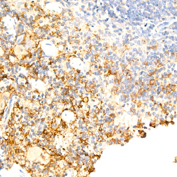
Immunohistochemistry analysis of paraffin-embedded Mouse spleen tissue using F4/80 Rabbit mAb (A23788) at a dilution of 1:500 (40x lens). High pressure antigen retrieval performed with 0.01M Tris-EDTA Buffer (pH 9.0) prior to IHC staining.
Immunohistochemistry analysis of paraffin-embedded Mouse liver tissue using F4/80 Rabbit mAb (A23788) at a dilution of 1:500 (40x lens). High pressure antigen retrieval performed with 0.01M Tris-EDTA Buffer (pH 9.0) prior to IHC staining.
Immunohistochemistry analysis of paraffin-embedded Mouse testis tissue using F4/80 Rabbit mAb (A23788) at a dilution of 1:500 (40x lens). High pressure antigen retrieval performed with 0.01M Tris-EDTA Buffer (pH 9.0) prior to IHC staining.
Immunohistochemistry analysis of paraffin-embedded Mouse thymus tissue using F4/80 Rabbit mAb (A23788) at a dilution of 1:500 (40x lens). High pressure antigen retrieval performed with 0.01M Tris-EDTA Buffer (pH 9.0) prior to IHC staining.
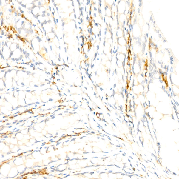
Immunohistochemistry analysis of paraffin-embedded Rat liver tissue using F4/80 Rabbit mAb (A23788) at a dilution of 1:500 (40x lens). High pressure antigen retrieval performed with 0.01M Tris-EDTA Buffer (pH 9.0) prior to IHC staining.
Immunohistochemistry analysis of paraffin-embedded Rat spleen tissue using F4/80 Rabbit mAb (A23788) at a dilution of 1:500 (40x lens). High pressure antigen retrieval performed with 0.01M Tris-EDTA Buffer (pH 9.0) prior to IHC staining.
Confocal imaging of paraffin-embedded Mouse spleen tissue using F4/80 Rabbit mAb (A23788, dilution 1:200) followed by a further incubation with Cy3 Goat Anti-Rabbit IgG (H+L) (AS007, dilution 1:500) (Red). DAPI was used for nuclear staining (Blue). High pressure antigen retrieval performed with 0.01M Citrate Buffer (pH 6.0) prior to IF staining. Objective: 40x.
Confocal imaging of paraffin-embedded Mouse liver tissue using F4/80 Rabbit mAb (A23788, dilution 1:200) followed by a further incubation with Cy3 Goat Anti-Rabbit IgG (H+L) (AS007, dilution 1:500) (Red). DAPI was used for nuclear staining (Blue). High pressure antigen retrieval performed with 0.01M Citrate Buffer (pH 6.0) prior to IF staining. Objective: 40x.
* VAT and and shipping costs not included. Errors and price changes excepted