
TOMM40 Rabbit mAb, Unconjugated, Monoclonal
Catalog Number:
ABB-A24644
- Images (9)

| Article Name: | TOMM40 Rabbit mAb, Unconjugated, Monoclonal |
| Biozol Catalog Number: | ABB-A24644 |
| Supplier Catalog Number: | A24644 |
| Alternative Catalog Number: | ABB-A24644-100UL,ABB-A24644-20UL |
| Manufacturer: | ABclonal |
| Host: | Rabbit |
| Category: | Antikörper |
| Application: | ELISA, IF, IHC-P, IP, WB |
| Species Reactivity: | Human |
| Immunogen: | Recombinant protein (or fragment).This information is considered to be commercially sensitive. |
| Conjugation: | Unconjugated |
| Alternative Names: | TOM40, PEREC1, C19orf1, PER-EC1, D19S1177E, TOMM40 |
| The protein encoded by this gene is localized in the outer membrane of the mitochondria. It is the channel-forming subunit of the translocase of the mitochondrial outer membrane (TOM) complex that is essential for import of protein precursors into mitochondria. Alternatively spliced transcript variants have been found for this gene. |
| Application Dilute: | WB,1:2000 - 1:10000|IP,0.5µg-4µg antibody for 400µg-600µg extracts of whole cells|IHC-P,1:100 - 1:500|IF/ICC,1:100 - 1:500|ELISA,Recommended starting concentration is 1 µg/mL. Please optimize the concentration based on your specific assay requirements. |
| Application Notes: | Cross-Reactivity: Human,Mouse,Rat. ResearchArea: Cancer,Signal Transduction,Endocrine Metabolism,Mitochondrial metabolism,Mitochondrial markers,Neuroscience,Neurodegenerative Diseases. Shipping: Ice Bag |
|
|
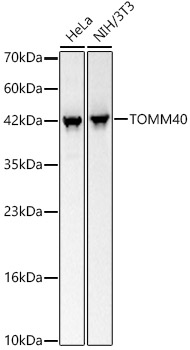
Western blot analysis of various lysates, using TOMM40 Rabbit mAb (A24644) at 1:4800 dilution. Secondary antibody: HRP-conjugated Goat anti-Rabbit IgG (H+L) (AS014) at 1:10000 dilution. Lysates/proteins: 25µg per lane. Blocking buffer: 3% nonfat dry milk in TBST. Detection: ECL Basic Kit (RM00020). Exposure time: 5s. |
|
|
Immunohistochemistry analysis of paraffin-embedded Human colon carcinoma using TOMM40 Rabbit mAb (A24644) at dilution of 1:400 (40x lens). High pressure antigen retrieval performed with 0.01M Citrate buffer (pH 6.0) prior to IHC staining. |
|
|
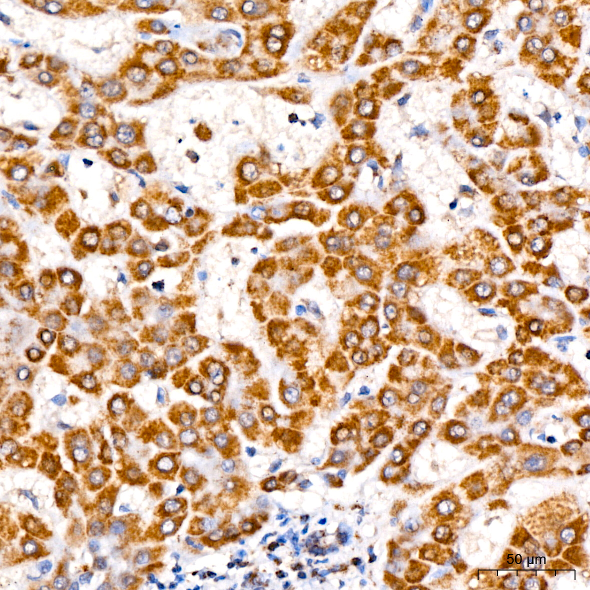
Immunohistochemistry analysis of paraffin-embedded Human liver using TOMM40 Rabbit mAb (A24644) at dilution of 1:400 (40x lens). High pressure antigen retrieval performed with 0.01M Citrate buffer (pH 6.0) prior to IHC staining. |
|
|
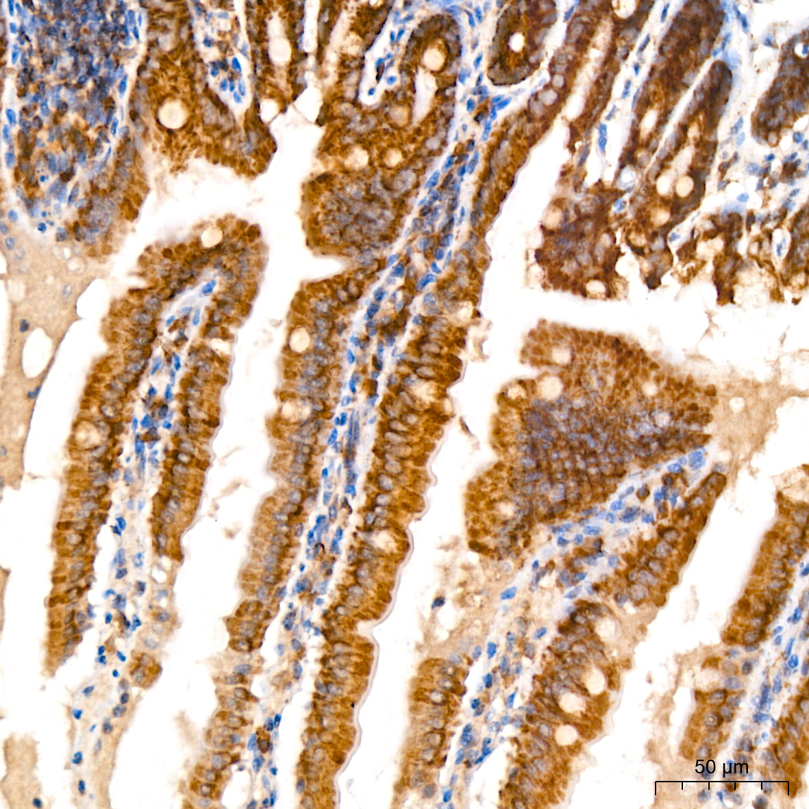
Immunohistochemistry analysis of paraffin-embedded Mouse colon using TOMM40 Rabbit mAb (A24644) at dilution of 1:400 (40x lens). High pressure antigen retrieval performed with 0.01M Citrate buffer (pH 6.0) prior to IHC staining. |
|
|
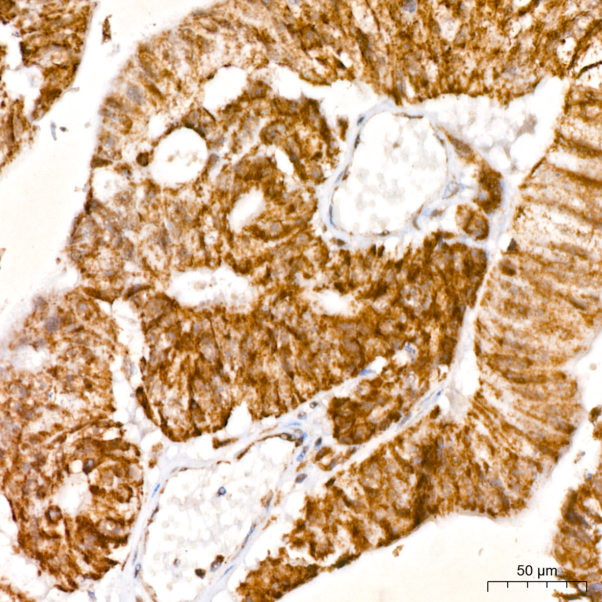
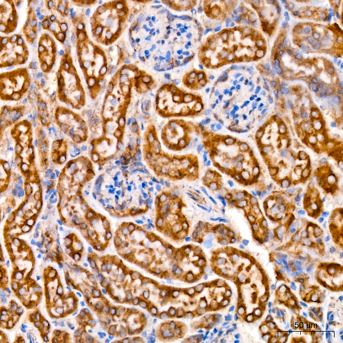
Immunohistochemistry analysis of paraffin-embedded Mouse kidney using TOMM40 Rabbit mAb (A24644) at dilution of 1:400 (40x lens). High pressure antigen retrieval performed with 0.01M Citrate buffer (pH 6.0) prior to IHC staining. |
|
|
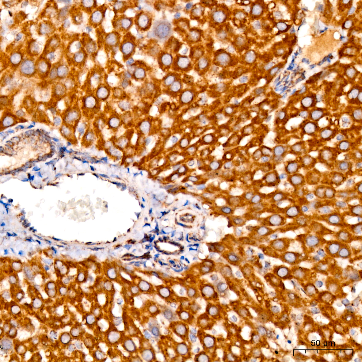
Immunohistochemistry analysis of paraffin-embedded Rat liver using TOMM40 Rabbit mAb (A24644) at dilution of 1:400 (40x lens). High pressure antigen retrieval performed with 0.01M Citrate buffer (pH 6.0) prior to IHC staining. |
|
|
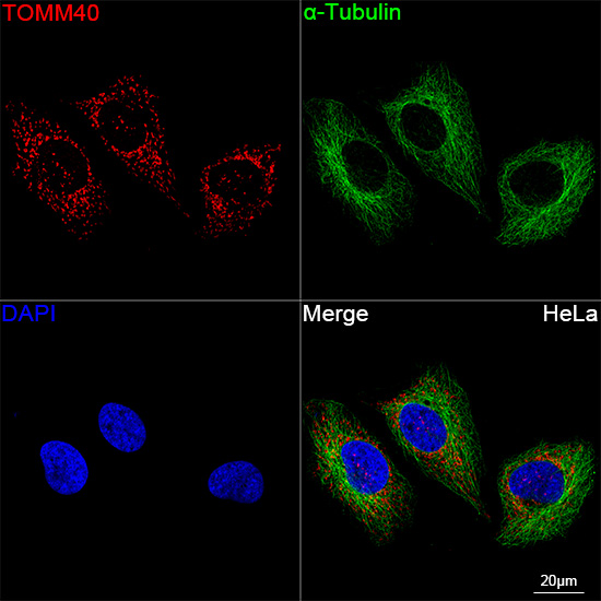
Confocal imaging of HeLa cells using TOMM40 Rabbit mAb (A24644,at dilution of 1:300) (red). The cells were counterstained with alpha-Tubulin Mouse mAb (AC012,dilution 1:400) (Green). DAPI was used for nuclear staining (blue). Objective: 100x. |
|
|
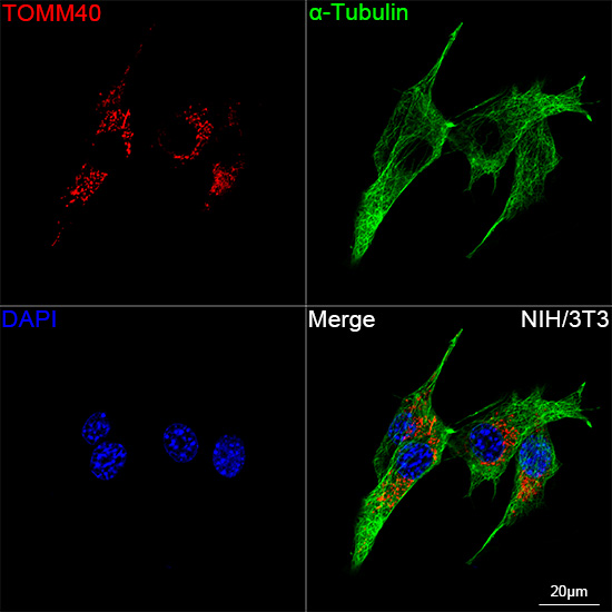
Confocal imaging of NIH/3T3 cells using TOMM40 Rabbit mAb (A24644,at dilution of 1:300) (Red). The cells were counterstained with alpha-Tubulin Mouse mAb (AC012,dilution 1:400) (Green). DAPI was used for nuclear staining (blue). Objective: 100x. |
|
|
Immunoprecipitation of TOMM40 from 500 µg extracts of Hep G2 cells was performed using 2 µg of TOMM40 Rabbit mAb (A24644). Rabbit Control IgG (AC005) was used to precipitate the Control IgG sample. IP samples were eluted with 1X Laemmli Buffer. The Input lane represents 10% of the total input. Western blot analysis of immunoprecipitates was conducted using TOMM40 Rabbit mAb (A24644) at a dilution of 1:5000. |
Product Guarantee and Expert Support
