
EEF1D Rabbit pAb, Unconjugated, Polyclonal
Catalog Number:
ABB-A2509
- Images (9)

| Article Name: | EEF1D Rabbit pAb, Unconjugated, Polyclonal |
| Biozol Catalog Number: | ABB-A2509 |
| Supplier Catalog Number: | A2509 |
| Alternative Catalog Number: | ABB-A2509-100UL,ABB-A2509-20UL,ABB-A2509-500UL,ABB-A2509-1000UL |
| Manufacturer: | ABclonal |
| Host: | Rabbit |
| Category: | Antikörper |
| Application: | ELISA, IF, IHC-P, IP, WB |
| Species Reactivity: | Human |
| Immunogen: | Recombinant protein (or fragment).This information is considered to be commercially sensitive. |
| Conjugation: | Unconjugated |
| Alternative Names: | EF1D, EF-1D, FP1047, EEF1D |
| This gene encodes a subunit of the elongation factor-1 complex, which is responsible for the enzymatic delivery of aminoacyl tRNAs to the ribosome. This subunit, delta, functions as guanine nucleotide exchange factor. It is reported that following HIV-1 infection, this subunit interacts with HIV-1 Tat. This interaction results in repression of translation of host cell proteins and enhanced translation of viral proteins. Several alternatively spliced transcript variants encoding multiple isoforms have been found for this gene. Related pseudogenes have been defined on chromosomes 1, 6, 7, 9, 11, 13, 17, 19. |
| Clonality: | Polyclonal |
| Molecular Weight: | 31kDa |
| NCBI: | 1936 |
| UniProt: | P29692 |
| Purity: | Affinity purification |
| Sequence: | MATNFLAHEKIWFDKFKYDDAERRFYEQMNGPVAGASRQENGASVILRDIARARENIQKSLAGSSGPGASSGTSGDHGELVVRIASLEVENQSLRGVVQELQQAISKLEARLNVLEKSSPGHRATAPQTQHVSPMRQVEPPAKKPATPAEDDEDDDIDLF |
| Target: | EEF1D |
| Antibody Type: | Primary Antibody |
| Application Dilute: | WB,1:500 - 1:2000|IHC-P,1:50 - 1:200|IF/ICC,1:50 - 1:200|IP,0.5µg-4µg antibody for 200µg-400µg extracts of whole cells|ELISA,Recommended starting concentration is 1 µg/mL. Please optimize the concentration based on your specific assay requirements. |
| Application Notes: | Cross-Reactivity: Human,Mouse,Rat. ResearchArea: Epigenetics Nuclear Signaling,Signal Transduction. Shipping: Ice Bag |
|
|
Immunohistochemistry analysis of paraffin-embeddedHuman liver tissue usingEEF1D Rabbit pAb(A2509) at a dilution of1:200 (40x lens).High pressure antigen retrieval was performed with 0.01 M citrate buffer (pH 6.0) prior to IHC staining. |
|
|
Immunohistochemistry analysis of paraffin-embeddedHuman kidney tissue usingEEF1D Rabbit pAb(A2509) at a dilution of1:200 (40x lens).High pressure antigen retrieval was performed with 0.01 M citrate buffer (pH 6.0) prior to IHC staining. |
|
|
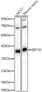
Western blot analysis of various lysates using EEF1D Rabbit pAb (A2509) at 1:2000 dilution. Secondary antibody: HRP-conjugated Goat anti-Rabbit IgG (H+L) (AS014) at 1:10000 dilution. Lysates / proteins: 25 µg per lane. Blocking buffer: 3 % nonfat dry milk in TBST. Detection: ECL Basic Kit (RM00020). Exposure time:10s. |
|
|
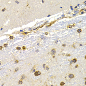
Immunohistochemistry analysis of paraffin-embeddedMouse brain tissue usingEEF1D Rabbit pAb(A2509) at a dilution of1:200 (40x lens).High pressure antigen retrieval was performed with 0.01 M citrate buffer (pH 6.0) prior to IHC staining. |
|
|
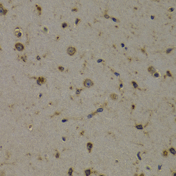
Immunohistochemistry analysis of paraffin-embeddedRat kidney tissue usingEEF1D Rabbit pAb(A2509) at a dilution of1:200 (40x lens).High pressure antigen retrieval was performed with 0.01 M citrate buffer (pH 6.0) prior to IHC staining. |
|
|
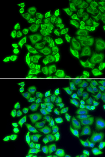
Immunofluorescence analysis of HeLa cells using EEF1D Rabbit pAb (A2509) at dilution of 1:200 (40x lens). Secondary antibody: Cy3-conjugated Goat anti-Rabbit IgG (H+L) (AS007) at 1:500 dilution. Blue: DAPI for nuclear staining. |
|
|
Immunofluorescence analysis of PC-12 cells using EEF1D Rabbit pAb (A2509) at dilution of 1:200 (40x lens). Secondary antibody: Cy3-conjugated Goat anti-Rabbit IgG (H+L) (AS007) at 1:500 dilution. Blue: DAPI for nuclear staining. |
|
|
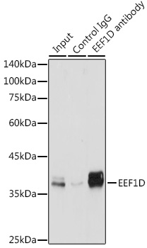
Immunoprecipitation of EEF1D in 300 µg extracts from jurkat cells using 3 µg EEF1D Rabbit pAb (A2509). Western blot analysis was performed using EEF1D Rabbit pAb (A2509) at 1:500 dilution. |
|
|
Immunoprecipitation of EEF1D in 300 µg extracts from Jurkat cells using 3 µg EEF1D Rabbit pAb (A2509). Western blot analysis was performed using EEF1D Rabbit pAb (A2509) at 1:500 dilution. |
Product Guarantee and Expert Support
