
hnRNP E2/PCBP2 Rabbit pAb, Unconjugated, Polyclonal
Catalog Number:
ABB-A2531
- Images (9)
| Article Name: | hnRNP E2/PCBP2 Rabbit pAb, Unconjugated, Polyclonal |
| Biozol Catalog Number: | ABB-A2531 |
| Supplier Catalog Number: | A2531 |
| Alternative Catalog Number: | ABB-A2531-20UL,ABB-A2531-100UL,ABB-A2531-500UL,ABB-A2531-1000UL |
| Manufacturer: | ABclonal |
| Host: | Rabbit |
| Category: | Antikörper |
| Application: | ELISA, IF, IHC-P, WB |
| Species Reactivity: | Human |
| Immunogen: | Synthetic peptide. This information is considered to be commercially sensitive. |
| Conjugation: | Unconjugated |
| Alternative Names: | HNRPE2, HNRNPE2, hnRNP-E2, hnRNP E2/PCBP2 |
| The protein encoded by this gene appears to be multifunctional. Along with PCBP-1 and hnRNPK, it is one of the major cellular poly(rC)-binding proteins. The encoded protein contains three K-homologous (KH) domains which may be involved in RNA binding. Together with PCBP-1, this protein also functions as a translational coactivator of poliovirus RNA via a sequence-specific interaction with stem-loop IV of the IRES, promoting poliovirus RNA replication by binding to its 5-terminal cloverleaf structure. It has also been implicated in translational control of the 15-lipoxygenase mRNA, human papillomavirus type 16 L2 mRNA, and hepatitis A virus RNA. The encoded protein is also suggested to play a part in formation of a sequence-specific alpha-globin mRNP complex which is associated with alpha-globin mRNA stability. This multiexon structural mRNA is thought to be retrotransposed to generate PCBP-1, an intronless gene with functions similar to that of PCBP2. This gene and PCBP-1 have paralogous genes (PCBP3 and PCBP4) which are thought to have arisen as a result of duplication events of entire genes. This gene also has two processed pseudogenes (PCBP2P1 and PCBP2P2). Multiple transcript variants encoding different isoforms have been found for this gene. |
| Clonality: | Polyclonal |
| Molecular Weight: | 39kDa |
| NCBI: | 5094 |
| UniProt: | Q15366 |
| Purity: | Affinity purification |
| Sequence: | GIPQSIIECVKQICVVMLETLSQSPPKGVTIPYRPKPSSSPVIFAGGQDRYSTGSDSASFPHTTPSMCLNPDLEGPPLEAYTIQGQYAIPQPDLTKLHQLAMQQSHFPMTHGNTGFSGIESSSPEVKGYWGLDASAQTTSHELTIPNDLIGCIIGRQGAKINEIRQMSGAQIKIANPVEGSTDRQVTITGSAASISLAQYLINVRLSSETGGMGSS |
| Target: | PCBP2 |
| Antibody Type: | Primary Antibody |
| Application Dilute: | WB,1:500 - 1:1000|IHC-P,1:50 - 1:200|IF/ICC,1:50 - 1:200|ELISA,Recommended starting concentration is 1 µg/mL. Please optimize the concentration based on your specific assay requirements. |
| Application Notes: | Cross-Reactivity: Human,Mouse,Rat. ResearchArea: Epigenetics Nuclear Signaling,RNA Binding. Shipping: Ice Bag |
|
|
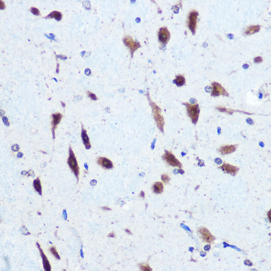
Immunohistochemistry analysis of paraffin-embedded Rat brain using hnRNP E2/PCBP2 Rabbit pAb (A2531) at dilution of 1:100 (40x lens). Microwave antigen retrieval performed with 0.01M Tris/EDTA Buffer (pH 9.0) prior to IHC staining. |
|
|
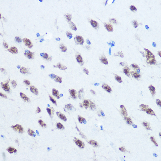
Immunohistochemistry analysis of paraffin-embedded Mouse brain using hnRNP E2/PCBP2 Rabbit pAb (A2531) at dilution of 1:100 (40x lens). Microwave antigen retrieval performed with 0.01M Tris/EDTA Buffer (pH 9.0) prior to IHC staining. |
|
|
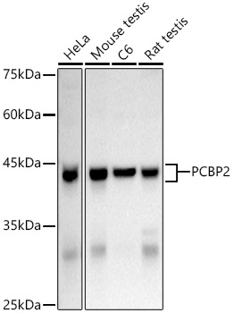
Western blot analysis of various lysates using hnRNP E2/PCBP2 Rabbit pAb (A2531) at 1:1000 dilution. Secondary antibody: HRP-conjugated Goat anti-Rabbit IgG (H+L) (AS014) at 1:10000 dilution. Lysates/proteins: 25µg per lane. Blocking buffer: 3% nonfat dry milk in TBST. Detection: ECL Basic Kit (RM00020). Exposure time: 10s. |
|
|
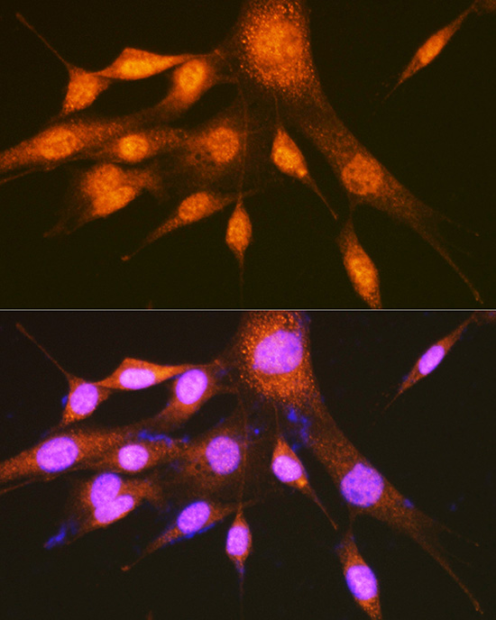
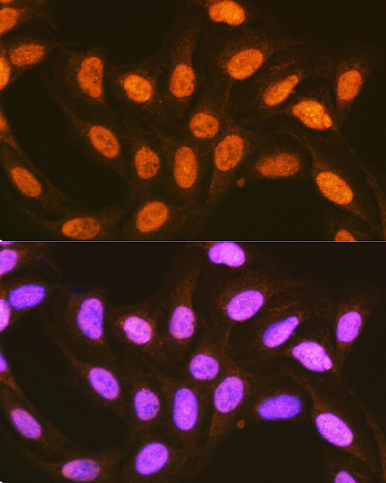
Immunofluorescence analysis of NIH-3T3 cells using hnRNP E2/PCBP2 Rabbit pAb (A2531) at dilution of 1:100 (40x lens). Secondary antibody: Cy3-conjugated Goat anti-Rabbit IgG (H+L) (AS007) at 1:500 dilution. Blue: DAPI for nuclear staining. |
|
|
Immunofluorescence analysis of U-2 OS cells using hnRNP E2/PCBP2 Rabbit pAb (A2531) at dilution of 1:100 (40x lens). Secondary antibody: Cy3-conjugated Goat anti-Rabbit IgG (H+L) (AS007) at 1:500 dilution. Blue: DAPI for nuclear staining. |
|
|
Immunofluorescence analysis of HeLa cells using hnRNP E2/PCBP2 Rabbit pAb (A2531) at dilution of 1:400 (40x lens). Secondary antibody: Cy3-conjugated Goat anti-Rabbit IgG (H+L) (AS007) at 1:500 dilution. Blue: DAPI for nuclear staining. |
|
|
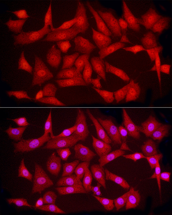
Immunofluorescence analysis of HepG2 cells using hnRNP E2/PCBP2 Rabbit pAb (A2531) at dilution of 1:400 (40x lens). Secondary antibody: Cy3-conjugated Goat anti-Rabbit IgG (H+L) (AS007) at 1:500 dilution. Blue: DAPI for nuclear staining. |
|
|
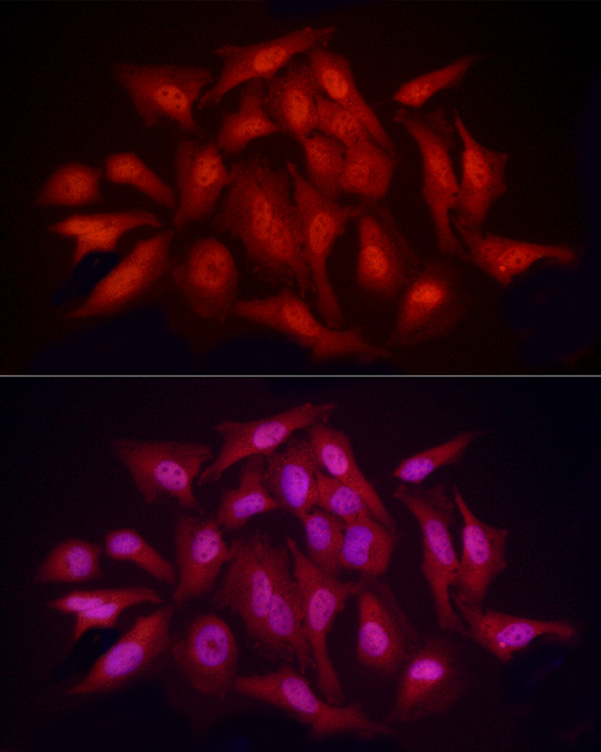
Immunofluorescence analysis of NIH/3T3 cells using hnRNP E2/PCBP2 Rabbit pAb (A2531) at dilution of 1:400 (40x lens). Secondary antibody: Cy3-conjugated Goat anti-Rabbit IgG (H+L) (AS007) at 1:500 dilution. Blue: DAPI for nuclear staining. |
|
|
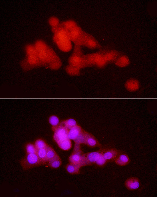
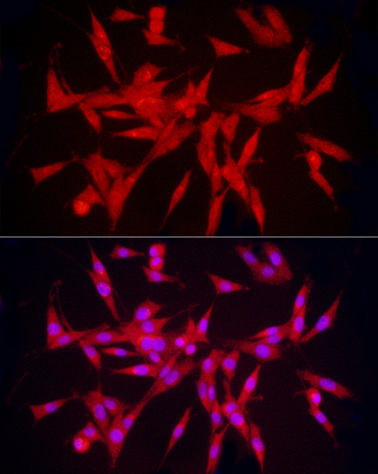
Immunofluorescence analysis of PC-12 cells using hnRNP E2/PCBP2 Rabbit pAb (A2531) at dilution of 1:400 (40x lens). Secondary antibody: Cy3-conjugated Goat anti-Rabbit IgG (H+L) (AS007) at 1:500 dilution. Blue: DAPI for nuclear staining. |
Product Guarantee and Expert Support
