
MAP2 Rabbit pAb, Unconjugated, Polyclonal
Catalog Number:
ABB-A2572
- Images (9)

| Article Name: | MAP2 Rabbit pAb, Unconjugated, Polyclonal |
| Biozol Catalog Number: | ABB-A2572 |
| Supplier Catalog Number: | A2572 |
| Alternative Catalog Number: | ABB-A2572-20UL,ABB-A2572-1000UL,ABB-A2572-100UL,ABB-A2572-500UL |
| Manufacturer: | ABclonal |
| Host: | Rabbit |
| Category: | Antikörper |
| Application: | ELISA, IF, IHC-P, WB |
| Species Reactivity: | Human |
| Immunogen: | Recombinant protein (or fragment).This information is considered to be commercially sensitive. |
| Conjugation: | Unconjugated |
| Alternative Names: | MAP-2, MAP2A, MAP2B, MAP2C, MAP2 |
| This gene encodes a protein that belongs to the microtubule-associated protein family. The proteins of this family are thought to be involved in microtubule assembly, which is an essential step in neurogenesis. The products of similar genes in rat and mouse are neuron-specific cytoskeletal proteins that are enriched in dentrites, implicating a role in determining and stabilizing dentritic shape during neuron development. A number of alternatively spliced variants encoding distinct isoforms have been described. |
| Clonality: | Polyclonal |
| Molecular Weight: | 200 kDa |
| NCBI: | 4133 |
| UniProt: | P11137 |
| Purity: | Affinity purification |
| Sequence: | GGESALAPSVFKQAKDKVSDGVTKSPEKRSSLPRPSSILPPRRGVSGDRDENSFSLNSSISSSARRTTRSEPIRRAGKSGTSTPTTPGSTAITPGTPPSYSSRTPGTPGTPSYPRTPHTPGTPKSAILVPSEKKVAIIRTP |
| Target: | MAP2 |
| Antibody Type: | Primary Antibody |
| Application Dilute: | WB,1:1000 - 1:5000|IF-P,1:50 - 1:200|IHC-P,1:50 - 1:200|ELISA,Recommended starting concentration is 1 µg/mL. Please optimize the concentration based on your specific assay requirements. |
| Application Notes: | Cross-Reactivity: Human,Mouse,Rat. ResearchArea: Signal Transduction,Cell Biology Developmental Biology,Cell Adhesion,Cytoskeleton,Microtubules,Neuroscience, Cell Type Marker,Stem Cells,Neuron marker. Shipping: Ice Bag |
|
|
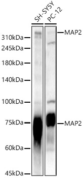
Western blot analysis of various lysates, using MAP2 Rabbit pAb (A2572) at 1:2000 dilution. Secondary antibody: HRP-conjugated Goat anti-Rabbit IgG (H+L) (AS014) at 1:10000 dilution. Lysates/proteins: 25µg per lane. Blocking buffer: 3% nonfat dry milk in TBST. Detection: ECL Basic Kit (RM00020). Exposure time: 10s. |
|
|
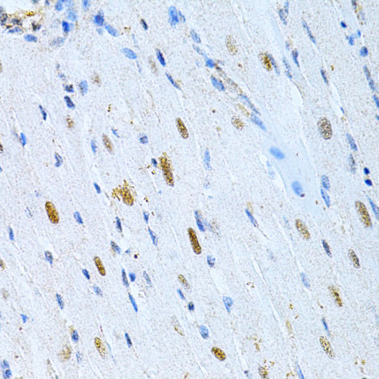
Immunohistochemistry analysis of paraffin-embedded Rat heart using MAP2 Rabbit pAb (A2572) at dilution of 1:200 (40x lens). Microwave antigen retrieval performed with 0.01M PBS Buffer (pH 7.2) prior to IHC staining. |
|
|
Western blot analysis of lysates from Mouse brain, using MAP2 Rabbit pAb (A2572) at 1:500 dilution. Secondary antibody: HRP-conjugated Goat anti-Rabbit IgG (H+L) (AS014) at 1:10000 dilution. Lysates/proteins: 25µg per lane. Blocking buffer: 3% nonfat dry milk in TBST. Detection: ECL Basic Kit (RM00020). Exposure time: 180s. |
|
|
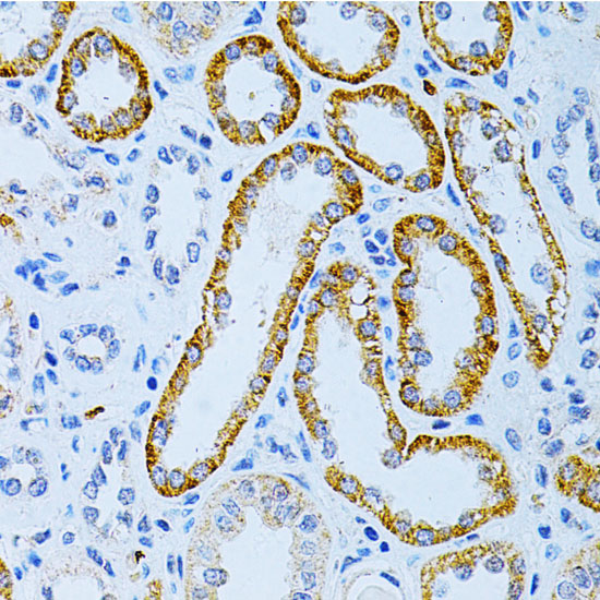
Immunohistochemistry analysis of paraffin-embedded Human kidney cancer using MAP2 Rabbit pAb (A2572) at dilution of 1:200 (40x lens). Microwave antigen retrieval performed with 0.01M PBS Buffer (pH 7.2) prior to IHC staining. |
|
|
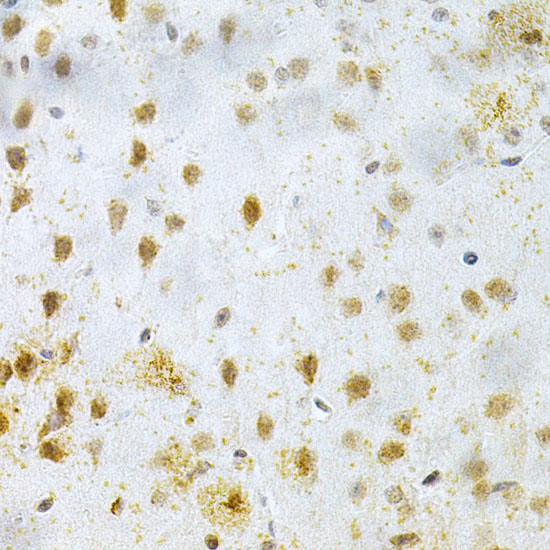
Immunohistochemistry analysis of paraffin-embedded Mouse brain using MAP2 Rabbit pAb (A2572) at dilution of 1:200 (40x lens). Microwave antigen retrieval performed with 0.01M PBS Buffer (pH 7.2) prior to IHC staining. |
|
|
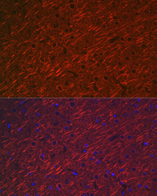
Immunofluorescence analysis of paraffin-embedded rat brain using MAP2 Rabbit pAb (A2572) at dilution of 1:100 (40x lens). Secondary antibody: Cy3-conjugated Goat anti-Rabbit IgG (H+L) (AS007) at 1:500 dilution. Blue: DAPI for nuclear staining. |
|
|
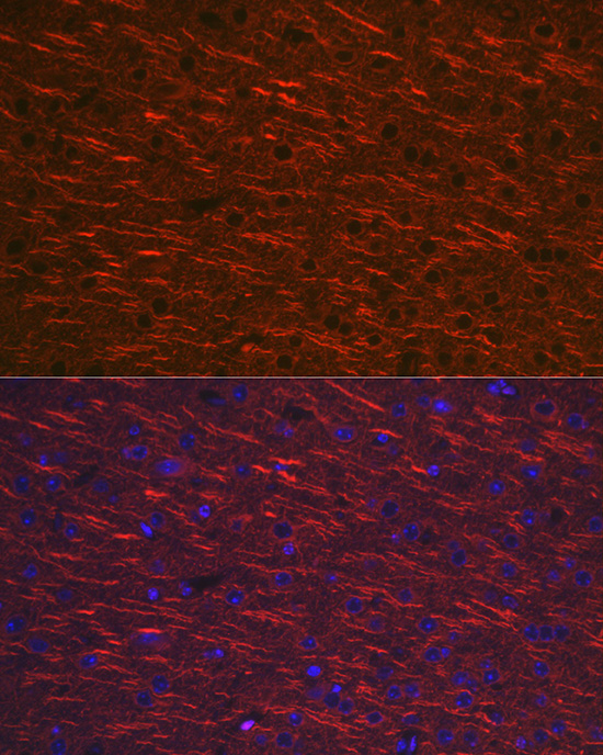
Immunofluorescence analysis of paraffin-embedded mouse brain using MAP2 Rabbit pAb (A2572) at dilution of 1:100 (40x lens). Secondary antibody: Cy3-conjugated Goat anti-Rabbit IgG (H+L) (AS007) at 1:500 dilution. Blue: DAPI for nuclear staining. |
|
|
Immunofluorescence analysis of paraffin-embedded mouse brain using MAP2 Rabbit pAb (A2572) at dilution of 1:20 (40x lens). Secondary antibody: Cy3-conjugated Goat anti-Rabbit IgG (H+L) (AS007) at 1:500 dilution. Blue: DAPI for nuclear staining. |
|
|
Immunofluorescence analysis of paraffin-embedded rat brain using MAP2 Rabbit pAb (A2572) at dilution of 1:20 (40x lens). Secondary antibody: Cy3-conjugated Goat anti-Rabbit IgG (H+L) (AS007) at 1:500 dilution. Blue: DAPI for nuclear staining. |
Product Guarantee and Expert Support
