
Phospho-FAK-Y576/577 Rabbit pAb, Unconjugated
Catalog Number:
ABB-AP0536
- Images (1)
| Article Name: | Phospho-FAK-Y576/577 Rabbit pAb, Unconjugated |
| Biozol Catalog Number: | ABB-AP0536 |
| Supplier Catalog Number: | AP0536 |
| Alternative Catalog Number: | ABB-AP0536-20UL, ABB-AP0536-100UL |
| Manufacturer: | ABclonal |
| Host: | Rabbit |
| Category: | Antikörper |
| Application: | ELISA, WB |
| Species Reactivity: | Human |
| Immunogen: | Synthetic peptide. This information is considered to be commercially sensitive. |
| Conjugation: | Unconjugated |
| Alternative Names: | FAK, FADK, FAK1, FRNK, FADK 1, PPP1R71, p125FAK, pp125FAK, Phospho-FAK-Y576/577 |
| This gene encodes a cytoplasmic protein tyrosine kinase which is found concentrated in the focal adhesions that form between cells growing in the presence of extracellular matrix constituents. The encoded protein is a member of the FAK subfamily of protein tyrosine kinases but lacks significant sequence similarity to kinases from other subfamilies. Activation of this gene may be an important early step in cell growth and intracellular signal transduction pathways triggered in response to certain neural peptides or to cell interactions with the extracellular matrix. Several transcript variants encoding different isoforms have been found for this gene. |
| Application Dilute: | WB,1:500 - 1:2000|ELISA,Recommended starting concentration is 1 µg/mL. Please optimize the concentration based on your specific assay requirements. |
| Application Notes: | Cross-Reactivity: Human, ResearchArea: Protein phosphorylation,Cancer,Signal Transduction,G protein signaling,G-Protein-Coupled Receptors Signaling to MAPK Erk,Kinase,Tyrosine kinases,PI3K-Akt Signaling Pathway,ErbB-HER Signaling Pathway,MAPK-Erk Signaling Pathway,Cell Biology Developmental Biology,Apoptosis,Cytoskeleton,Actins,Extracellular Matrix,Immunology Inflammation,Cardiovascular,Angiogenesis. |
|
|
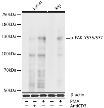
Western blot analysis of various lysates, using Phospho-FAK-Y576/577 Rabbit pAb (AP0536) at 1:1000 dilution. Jurkat cells were treated with CD-3 (10 µg/mL) for 2 minutes after serum-starvation overnight or treated with PMA/TPA (200nM) for 10 minutes. Raji cells were treated with PMA/TPA (200nM) for 30 minutes. Secondary antibody: HRP-conjugated Goat anti-Rabbit IgG (H+L) (AS014) at 1:10000 dilution. Lysates/proteins: 25µg per lane. Blocking buffer: 3% BSA. Detection: ECL Basic Kit (RM00020). Exposure time: 90s. |
Product Guarantee and Expert Support
