
Phospho-p53-S367 Rabbit pAb
Catalog Number:
ABB-AP1170
- Images (1)
| Article Name: | Phospho-p53-S367 Rabbit pAb |
| Biozol Catalog Number: | ABB-AP1170 |
| Supplier Catalog Number: | AP1170 |
| Alternative Catalog Number: | ABB-AP1170-20UL, ABB-AP1170-100UL |
| Manufacturer: | ABclonal |
| Host: | Rabbit |
| Category: | Antikörper |
| Application: | ELISA, WB |
| Species Reactivity: | Human |
| Immunogen: | Synthetic peptide. This information is considered to be commercially sensitive. |
| Alternative Names: | P53, BCC7, LFS1, BMFS5, TRP53, Phospho-p53-S367 |
| This gene encodes a tumor suppressor protein containing transcriptional activation, DNA binding, and oligomerization domains. The encoded protein responds to diverse cellular stresses to regulate expression of target genes, thereby inducing cell cycle arrest, apoptosis, senescence, DNA repair, or changes in metabolism. Mutations in this gene are associated with a variety of human cancers, including hereditary cancers such as Li-Fraumeni syndrome. Alternative splicing of this gene and the use of alternate promoters result in multiple transcript variants and isoforms. Additional isoforms have also been shown to result from the use of alternate translation initiation codons from identical transcript variants (PMIDs: 12032546, 20937277). |
| Application Dilute: | WB,1:100 - 1:500|ELISA,Recommended starting concentration is 1 µg/mL. Please optimize the concentration based on your specific assay requirements. |
| Application Notes: | Cross-Reactivity: Human, ResearchArea: Epigenetics Nuclear Signaling,Transcription Factors,DNA Damage Repair,Protein phosphorylation,Cancer,Tumor suppressors,p53 pathway,Signal Transduction,PI3K-Akt Signaling Pathway,ErbB-HER Signaling Pathway,MAPK-JNK Signaling Pathway,ATM Signaling Pathway,Cell Biology Developmental Biology,Apoptosis,Mitochondrial Control of Apoptosis,Cell Cycle,Cell cycle inhibitors,Cell Cycle Control-G1 S Checkpoint,Cell Cycle Control-G2 M DNA Damage Checkpoint,Endocrine Metabolism,AMPK Signaling Pathway,Warburg Effect,Neuroscience,Neurodegenerative Diseases. |
|
|
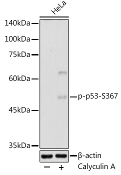
Western blot analysis of lysates from HeLa cells, using Phospho-p53-S367 Rabbit pAb (AP1170) at 1:500 dilution. HeLa cells were treated with Calyculin A (100 nM) at 37°C for 30 minutes after serum-starvation overnight. Secondary antibody: HRP-conjugated Goat anti-Rabbit IgG (H+L) (AS014) at 1:10000 dilution. Lysates/proteins: 25µg per lane. Blocking buffer: 3% nonfat dry milk in TBST. Detection: ECL Enhanced Kit (RM00021). Exposure time: 180s. |
Product Guarantee and Expert Support
