A synthetic peptide corresponding to amino acids 41-54 (GAAPAPGIFSSQPG-Cys), of human Bcl-2 was used as immunogen.
Alternative Names:
BCL2
Bcl-2 (B-cell lymphoma 2), is a member of Bcl-2 family of regulator proteins. These proteins contain a hydrophobic cleft that binds to BH3-only proteins and to the pro-apoptotic Bcl-2 family members Bad, Bak, and Bax to inhibit apoptosis. In the absence of this binding, the proapoptotic Bcl-2 members are recruited to the OMM (Outer Mitochondrial Membrane) at which they oligomerize and cause OMM permeabilization, releasing proapoptotic effectors such as SMAC or cytochrome-c. Bcl-2 also neutralize a group of sensor proteins (such as BIM), which are triggered by cytotoxic stimuli such as chemotherapy. BCL-2 proteins therefore have a central role as guardians against apoptosis, helping cancer cells to evade cell death.
Western blot analysis: 2-4 µg/ml, Immunohistochemical analysis: 5 µg/ml, Flowcytometric analysis- 2-4 µg/10 6 Cells
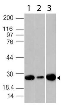
Figure-1: Western blot analysis of Bcl2. Anti- Bcl2(Clone: BC1) was used at 2 µg/ml on 293, MCF7 and Jurkat lysates.
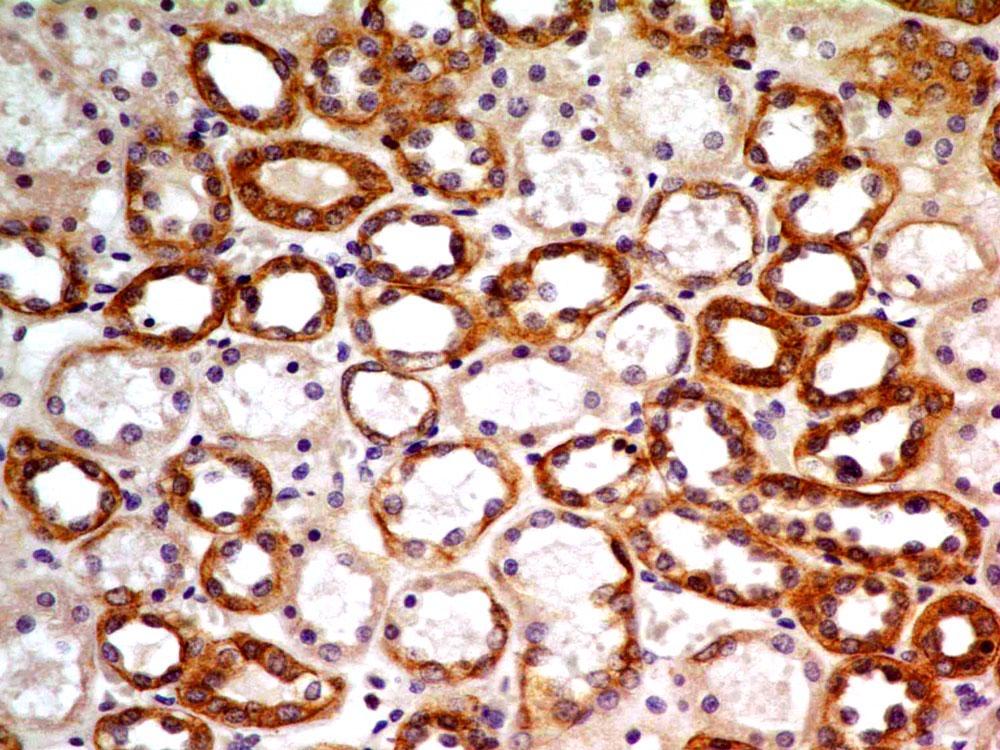
Figure-2 : Immunohistochemical analysis of Bcl-2 in human kidney tissue using Bcl-2 antibody (Clone: BC1) at 5 µg/ml.
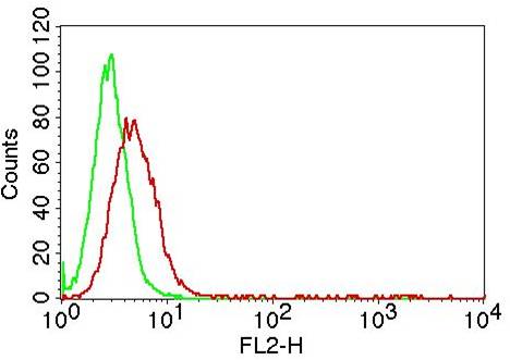
Figure-6: Intracellular flow cytometric analysis of Bcl-2 in Jurkat cell lines using 2 µg/10 6 cells of Anti-Bcl-2 antibody (10-1052 Abeomics) . Green represent isotype control and red represent Anti-Bcl-2 antibody (Clone: BC1). Goat anti-mouse PE conjugated secondary antibody (ABEOMICS) was used.
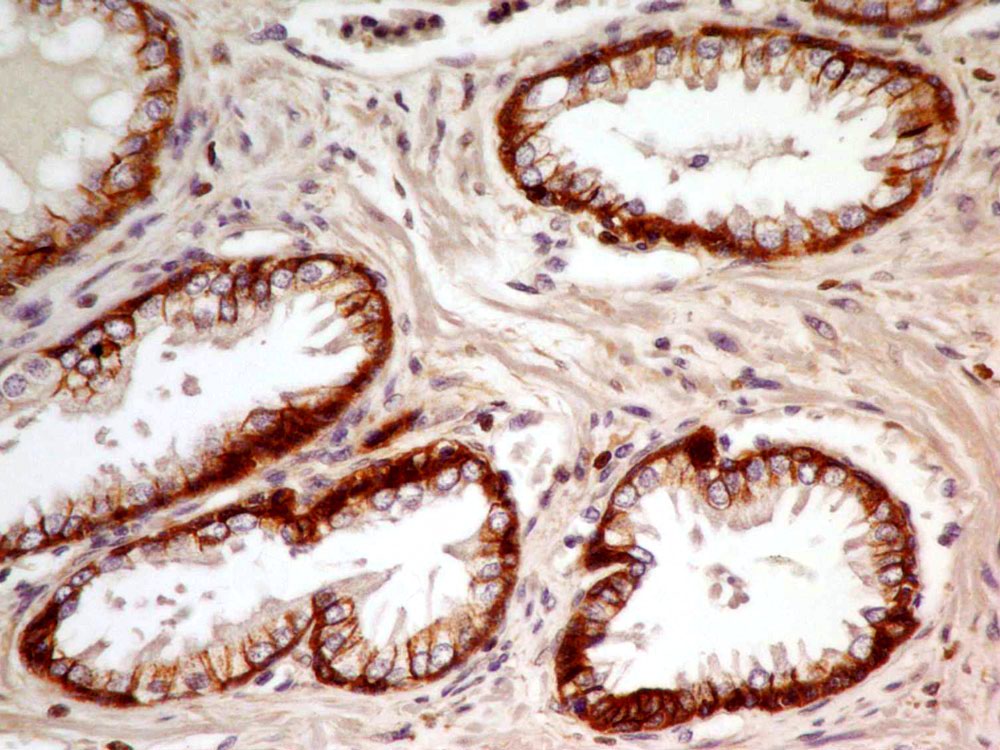
Figure-3 : Immunohistochemical analysis of Bcl-2 in human Prostate tissue using Bcl-2 antibody (Clone: BC1) at 5 µg/ml.
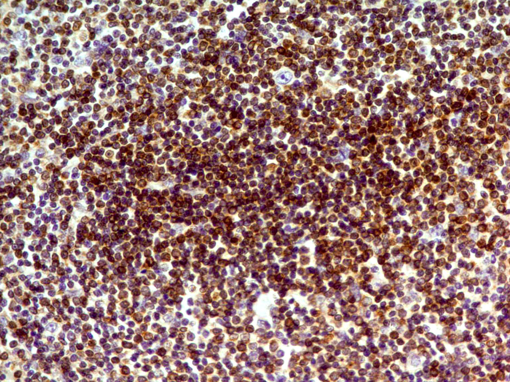
Figure-4 : Immunohistochemical analysis of Bcl-2 in human Spleen tissue using Bcl-2 antibody (Clone: BC1) at 5 µg/ml.
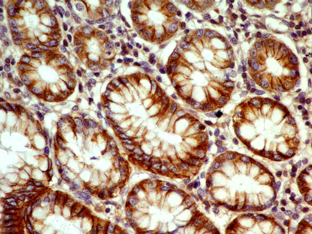
Figure-5 : Immunohistochemical analysis of Bcl-2 in human Stomach tissue using Bcl-2 antibody (Clone: BC1) at 5 µg/ml.
* VAT and and shipping costs not included. Errors and price changes excepted