Full length recombinant protein of BST 2 was used as the immunogen for this antibody.
Alternative Names:
Bone marrow stromal antigen 2, HM1.24 antigen, Tetherin, CD317
BST2 (Bone Marrow Stromal Antigen 2), a type II transmembrane protein also known as HM1.24/CD317, has been identified to be overexpressed in a variety of cell lines from different cancer types, including multiple myeloma, breast, lung, and kidney cancers. BST2 was discovered as a marker of differentiated B cells and was later rediscovered as a potent antiviral restriction factor with the ability to tether enveloped viruses to the cell membrane of infected cells via its GPI anchor as well as to potently inhibit virus replication in cultured cells and in vivo. It also acts as an innate immune sensor of HIV-1 that elicits an inflammatory response upon exposure. BST2 mediate host immune response by activating NF-KB through interaction with TAK1 (Transforming Growth Factor Beta-Activated Kinase 1) and TNF receptor associated factors (TRAFs) 2 and 6. In addition, BST2 induces ADCC (antibody-dependent cell cytotoxicity) against the envelope protein of HIV. BST2 was expressed constitutively on the cell surface of both CD3 and CD3+ thymocytes ex vivo.
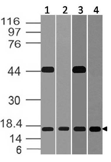
Fig-4: Western blot analysis of BST 2. Anti-BST 2 antibody (Clone: ABM52D8) was used at 2 µg/ml on (1) m Lung, (2) r Lung, (3) m Kidney and (4) r Kidney lysates.
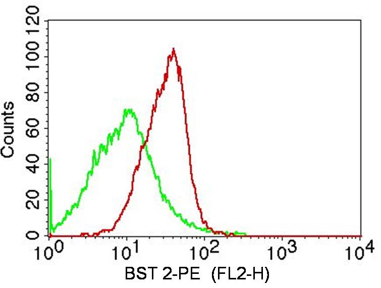
Fig-1: Cell surface flow analysis of BST2 in PBMCs (lymphocytes gated) using 0.5 µg/10 6 cells of antibody (Clone: ABM52D8). Green represents isotype control, red represents anti-BST2 antibody. Goat anti-mouse PE conjugate was used as secondary antibody. (Cells were incubated with primary antibody for 45 min. then washed twice with PBS by centrifuging at 1100 rpm for 5 min, followed by 30 min incubation with conjugated secondary antibody. Data acquisition was done after washing twice with PBS as
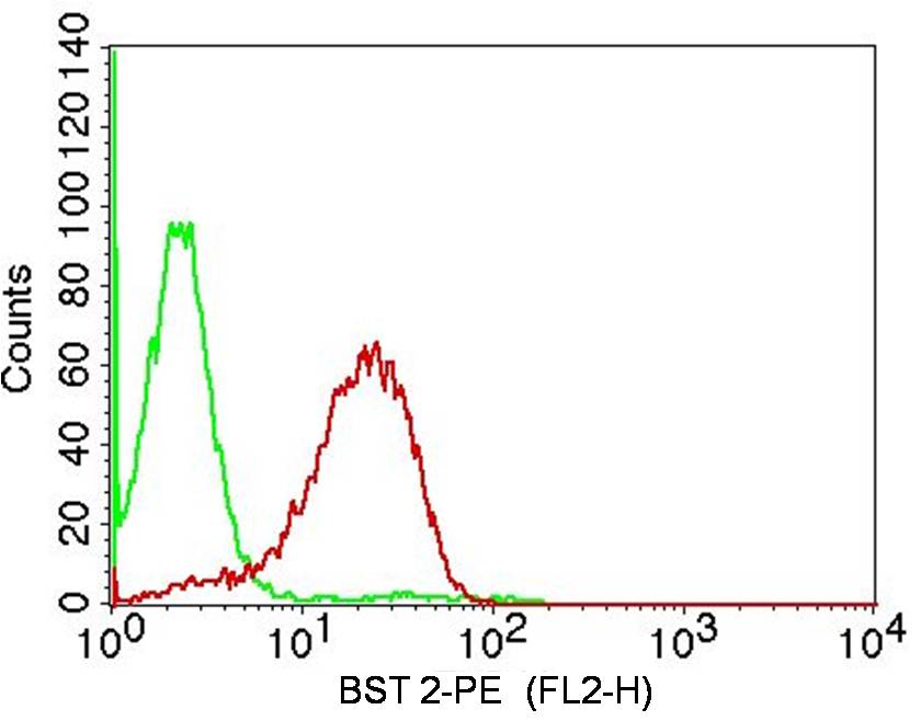
Fig-2: Cell surface flow analysis of BST2 on Molt4 cells using 0.5 µg/10 6 cells of antibody (Clone: ABM52D8). Green represents isotype control, red represents anti-BST2 antibody. Goat anti-mouse PE conjugate was used as secondary antibody. (Cells were incubated with primary antibody for 45 min. then washed twice with PBS by centrifuging at 1100 rpm for 5 min, followed by 30 min incubation with conjugated secondary antibody. Data acquisition was done after washing twice with PBS as mentioned a
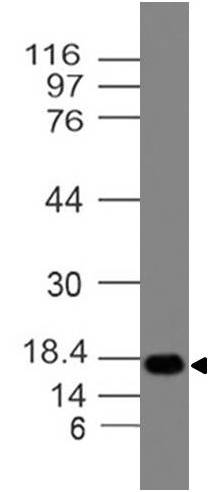
Fig-3: Western blot analysis of BST 2. Anti-BST 2 antibody (Clone: ABM52D8) was used at 2 µg/ml on Ramos lysate.
* VAT and and shipping costs not included. Errors and price changes excepted