A partial length recombinant Trim29 protein (amino acids 2-291) was used as the immunogen for this antibody.
Alternative Names:
TRIM29||ATDC
TRIM29 (Tripartite motif-containing 29) is a member of the tripartite motif (TRIM) family of transcription factors, characterized by the conserved RING finger, B-box, and coiled-coil domains. The TRIM family has been implicated in a variety of physiologic processes including development, apoptosis and epithelial-mesenchymal transition (EMT). Depending on a tumor types, TRIM29 also may function as an oncogene or a tumor suppressor. As an oncogene, it facilitates tumor cell proliferation and invasion through stabilization of Beta-catenin. The mechanisms may also involve deactivation of p53 activity and promoting cell survival by inhibiting proapoptotic genes regulated by p53. As a histone binding protein TRIM29 is responsible for DDR (DNA damage response). It functions as a scaffold protein to assemble DNA repair proteins into chromatin followed by efficient activation of DDR. TRIM29 binds to modified histone H3 and H4 tails in the context of nucleosomes. Furthermore, chromatin binding of TRIM29 is required for the phosphorylation of H2AX nucleosomes and cell viability in response to ionizing radiation.
Western blot analysis: 1-2 µg/ml, Immunohistochemical analysis: 1 µg/ml
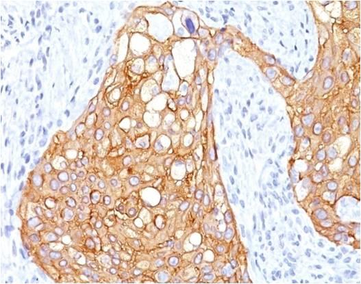
Figure-2 : Immunohistochemical analysis of Trim29 in human lung adenocarcinoma tissue using Trim29 antibody (Clone: ABM43D2) at 1 µg/ml.
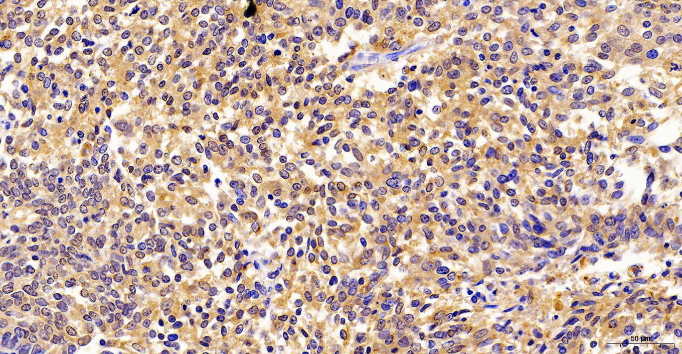
Figure-3 : Immunohistochemical analysis of Trim29in human Lungs squamous cell carcinoma tissue (20X) using Trim29 antibody (Clone: ABM43D2).
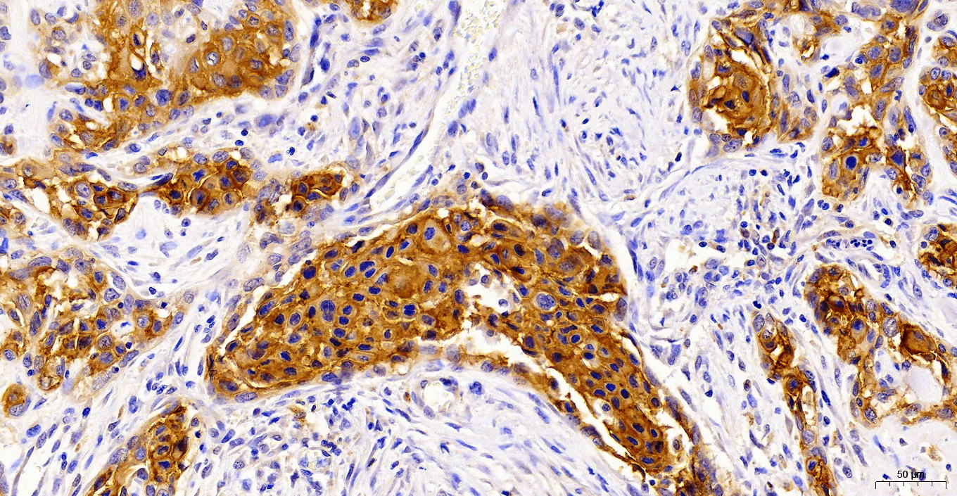
Figure-4 : Immunohistochemical analysis of Trim29in Esophagus squamous cell carcinoma tissue (20X) using Trim29 antibody (Clone: ABM43D2).
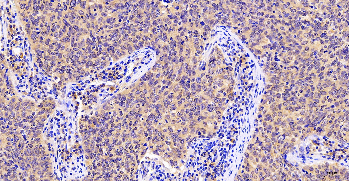
Figure-5 : Immunohistochemical analysis of Trim29in Renal cell carcinoma tissue (20X) using Trim29 antibody (Clone: ABM43D2) .
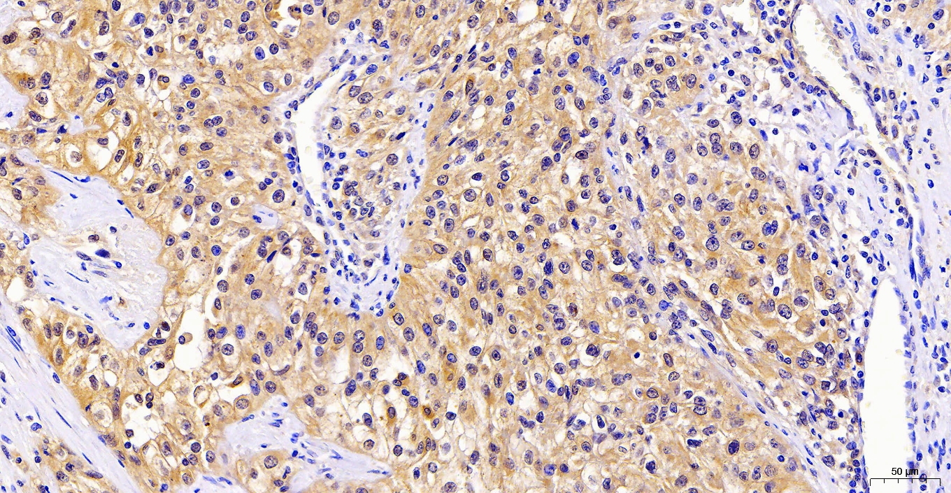
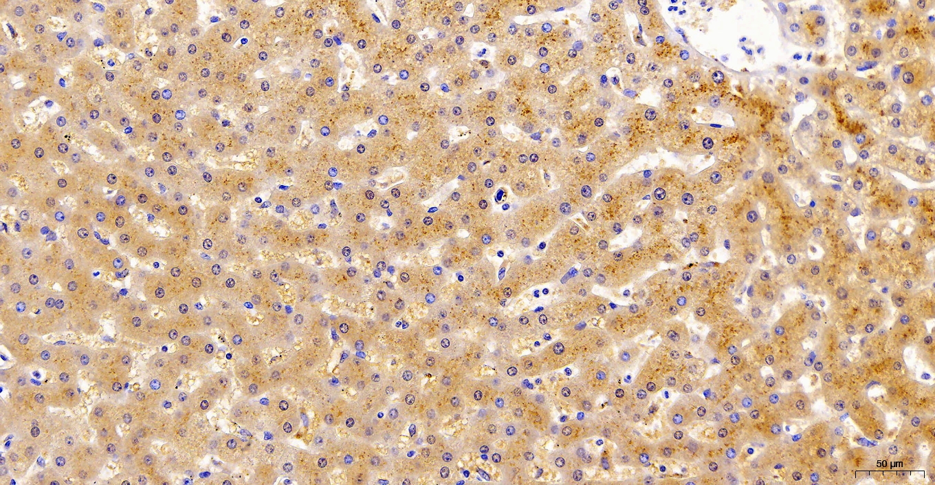
Figure-6 : Immunohistochemical analysis of Trim29in Hepatocellular carcinoma tissue (20X) using Trim29 antibody (Clone: ABM43D2).
Figure-7 : Immunohistochemical analysis of Trim29in Uterus endometrial carcinoma tissue (20X) using Trim29 antibody (Clone: ABM43D2).
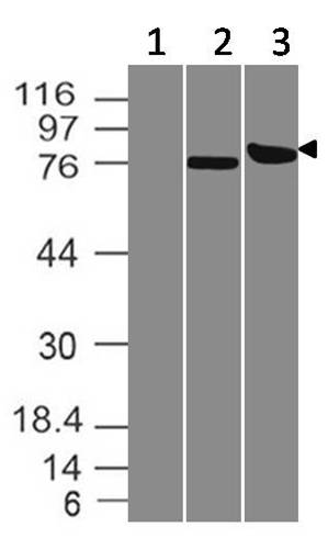
Figure-1: Western blot analysis of Trim29. Anti- Trim29 antibody (Clone: ABM43D2) was tested at 2 µg/ml on (1) HEK 293 cell lysate, (2) A431 cell lysate and (3) recombinant Trim29 protein expressed in HEK 293 cells.
* VAT and and shipping costs not included. Errors and price changes excepted