Monoclonal antibody to Human PD-L1 (Clone: ABM5F25)
Biozol Catalog Number:
ABI-10-7599-25
Supplier Catalog Number:
10-7599-25
Alternative Catalog Number:
ABI-10-7599-25-25UG
Manufacturer:
Abeomics
Host:
Mouse
Category:
Antikörper
Application:
FACS, IHC, WB
Species Reactivity:
Human
Immunogen:
A partial length recombinant protein of PD-L1 (amino acid 13-224) was used as the immunogen for this antibody.
Alternative Names:
CD274||B7H1||PDCD1L1||PDCD1LG1||PDL1
PD-L1 (CD274/B7-H1) is a critical membrane-bound costimulatory molecule belonging to the B7 superfamily that inhibits immune responses through its receptor, PD-1. PD-L1 plays a key role in the pathogenesis of inflammatory diseases (programmed death 1). It is widely expressed in the mononuclear phagocyte system (MPS), may co-stimulate T cells, and regulates inflammatory responses. PD-L1 exerts inflammation regulatory functions via a negative co-stimulatory effect on T cell functions to inhibit cytokine secretion, facilitates apoptosis of activated T cells, and induces T cell anergy. Aberrant expression and dysregulation of CD274 have been reported during bacterial infection, inflammation, and in numerous autoimmune diseases.
FACS analysis: 0.5-1 µg/10 6 cells, Western blot analysis: 2-4 µg/ml, Immunohistochemical analysis: 5-10 µg/ml
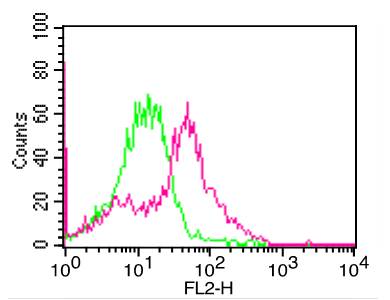
Fig-2: Cell surface flow analysis of PD-L1 in CHO-PD-L1 transfected cell line using 0.5 µg/10 6 cells of PD-L1 antibody (Clone: ABM5F25). Green represents isotype control, red represents anti-PD-L1 antibody. Goat anti-mouse PE conjugate was used as secondary antibody.
Fig:1- Cell Surface flow analysis of PD-L1 in 3 day-PHA treated human PBMC cells using 1 µg/10 6 cells of PD-L1 antibody (Clone: ABM5F25). Green represents isotype control, red represents anti-PD-L1 antibody. Goat anti-mouse PE conjugate was used as secondary antibody.
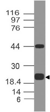
Fig-3: Western blot analysis of PDL1. Anti-PD-L1 antibody (Clone: ABM5F25) was tested at 0.5 µg/ml on Recombinant lysate.
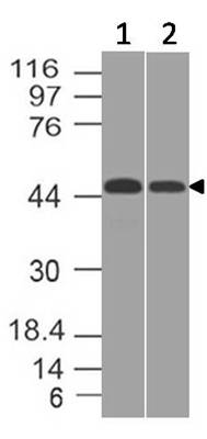
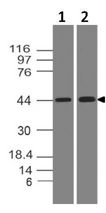
Fig-6: Western blot analysis of PDL1. Anti-PD-L1 antibody (Clone: ABM5F25) was tested at 0.5 µg/ml on (1) U87 and (2) THP1 lysates.
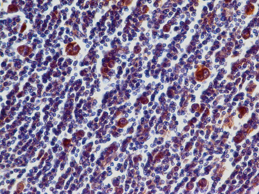
Fig-5: Immunohistochemical analysis of PD-L1 in Hodkins Lymphoma tissue using PD-L1 antibody (Clone: ABM5F25) at 5 µg/ml.
Fig-4: Western blot analysis of PDL1. Anti-PD-L1 antibody (Clone: ABM5F25) was tested at 2 µg/ml on (1) Daudi and (2) HepG2 lysates.
* VAT and and shipping costs not included. Errors and price changes excepted