Western blot analysis: 2-4 µg/ml, FACS analysis-0.5-1 µg/10 6 Cells, Immunohistochemical analysis-2-4 µg/ml
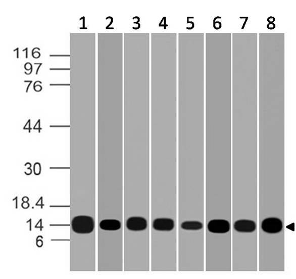
Figure:1- Western blot analysis of Galectin-1. Anti-Galectin-1 antibody (Clone: ABM5B54) was tested at 0.01 µg/ml on (1) Recombinant protein and 2 µg/ml on (2) Hela, (3) 3T3, (4) PC3, (5) 293, (6) THP1, (7) K562 and (8) HCT-116 lysates.
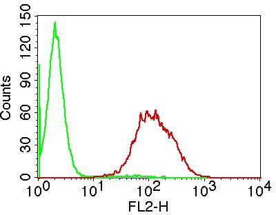
Figure:2- Intracellular flow analysis of Galectin-1 in U87 cell line using 0.5 µg/10 6 cells of Galectin-1 antibody (Clone: ABM5B54). Green represents isotype control, red represents anti-Galectin-1 antibody (10-7618). Goat anti-mouse PE conjugate was used as secondary antibody.
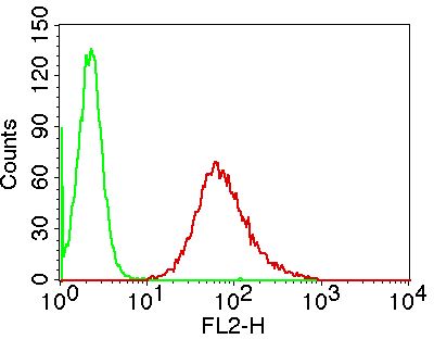
Figure:3- Intracellular flow analysis of Galectin-1 in A431 cell line using 0.5 µg/10 6 cells of Galectin-1 antibody (Clone: ABM5B54). Green represents isotype control, red represents anti-Galectin-1 antibody (10-7618). Goat anti-mouse PE conjugate was used as secondary antibody.
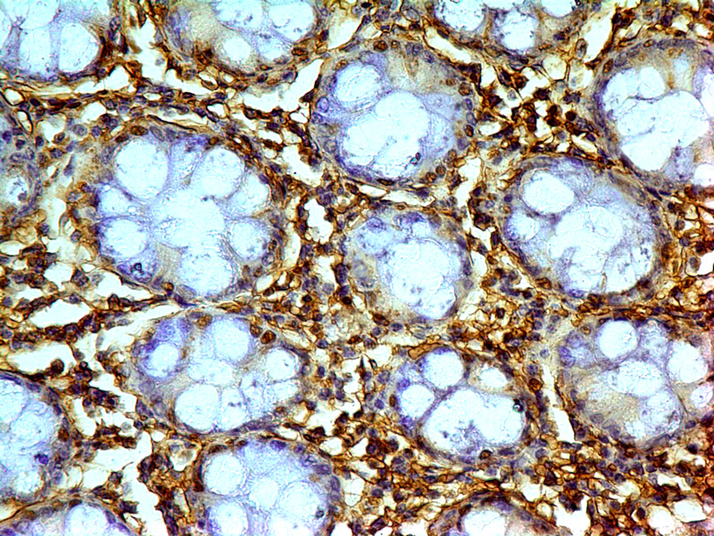
Figure:4-Immunohistochemical analysis of Galectin-1. Anti-Galectin-1 (Clone: ABM5B54) was used in Colon Carcinoma tissue at 2 µg/ml.
* VAT and and shipping costs not included. Errors and price changes excepted