
Monoclonal Antibody to CD137 / 4-1BB (Clone: ABM3D3.2E8)
Catalog Number:
ABI-10-7620
- Images (4)
| Article Name: | Monoclonal Antibody to CD137 / 4-1BB (Clone: ABM3D3.2E8) |
| Biozol Catalog Number: | ABI-10-7620 |
| Supplier Catalog Number: | 10-7620 |
| Alternative Catalog Number: | ABI-10-7620-100UG |
| Manufacturer: | Abeomics |
| Host: | Mouse |
| Category: | Antikörper |
| Application: | FACS, IHC, WB |
| Species Reactivity: | Human |
| Immunogen: | A partial length recombinant protein of CD137 was used as the immunogen for this antibody. |
| Alternative Names: | Tumor necrosis factor receptor superfamily member 9, 4-1BB ligand receptor, T-cell antigen 4-1BB homolog, T-cell antigen ILA |
| Application Dilute: | Western blot analysis: 4-6 µg/ml, Immunohistochemical Analysis: 2-4 µg/ml, Facs anaysis: 1-2 µg/10 6 Cells |
|
|
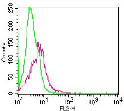
Figure-1: Cell surface staining of PHA stimulated PBMC. Green represents isotype control, red represents Anti-CD137 (10-7620). 1 µg antibody was used. Goat anti-mouse PE was used as secondary antibody. |
|
|
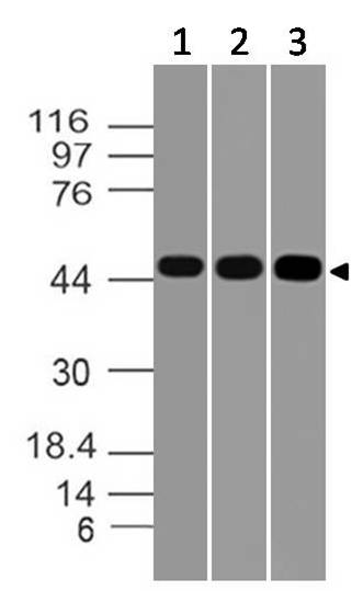
Figure-4: Westernblot analysis of CD137. Anti- CD137 (Clone: ABM3D3.2E8) was tested at 4 µg/ml on Jurkat, HepG2 and Molt-4. |
|
|
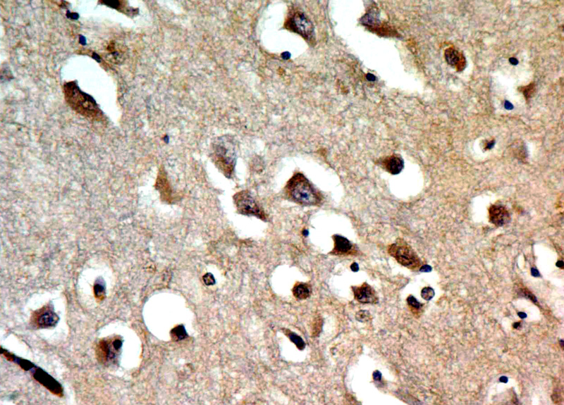
Figure-2: Immunohistochemical analysis of CD137. Anti-CD137 (Clone: ABM3D3.2E8) was used in Human Brain tissue at 2 µg/ml. |
|
|
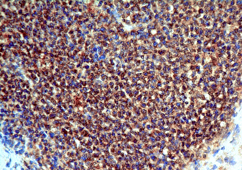
Figure-3: Immunohistochemical analysis of CD137. Anti-CD137 (Clone: ABM3D3.2E8) was used in Human Tonsil tissue at 2 µg/ml. |
Product Guarantee and Expert Support
