Western blot analysis: 4-6 µg/ml, Facs anaysis: 1-2 µg/10 6 Cells
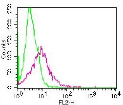
Figure-1: Cell surface staining of PHA stimulated PBMC. Green represents isotype control, red represents Anti-CD137 (10-7621). 1 µgantibody was used. Goat anti-mouse PE was used as secondary antibody.
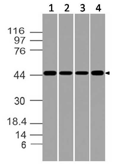
Figure-2: Western blot analysis of CD137. Anti- CD137 (Clone: ABM2B2.1A5) was tested at 4 µg/ml on (1) Jurkat, (2) MOLT-4, (3) K562 and (4) HepG2 lysates.
* VAT and and shipping costs not included. Errors and price changes excepted