
Anti-HDAC1 Recombinant Antibody, IgG1, Clone: [CL0510], Mouse, Monoclonal
Catalog Number:
ATA-AMAB90781R
- Images (8)

| Article Name: | Anti-HDAC1 Recombinant Antibody, IgG1, Clone: [CL0510], Mouse, Monoclonal |
| Biozol Catalog Number: | ATA-AMAB90781R |
| Supplier Catalog Number: | AMAb90781R |
| Alternative Catalog Number: | ATA-AMAB90781R-100,ATA-AMAB90781R-25 |
| Manufacturer: | Atlas Antibodies |
| Host: | Mouse |
| Category: | Antikörper |
| Application: | ICC, IHC, WB |
| Species Reactivity: | Human, Mouse, Rat |
| Alternative Names: | GON-10, HD1, RPD3L1 |
| Recombinant Mouse Monoclonal Anti-HDAC1 Antibody against Human histone deacetylase 1. Validated for Immunofluorescence, Immunohistochemistry and Western Blot. |
|
|
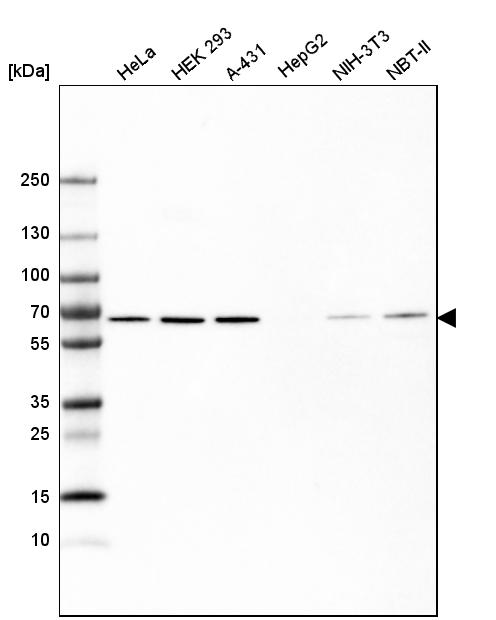
Western blot analysis in human cell line HeLa, human cell line HEK 293, human cell line A-431, human cell line HepG2, mouse cell line NIH-3T3 and rat cell line NBT-II. |
|
|
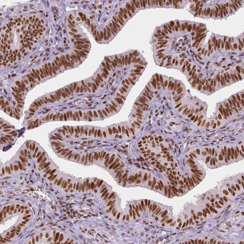
Immunohistochemical staining of human fallopian tube shows strong nuclear positivity in glandular cells. |
|
|
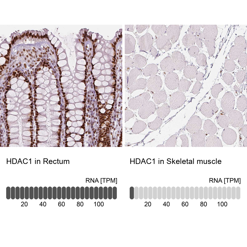
Immunohistochemistry analysis in human rectum and skeletal muscle tissues using AMAb90781 antibody. Corresponding HDAC1 RNA-seq data are presented for the same tissues. |
|
|
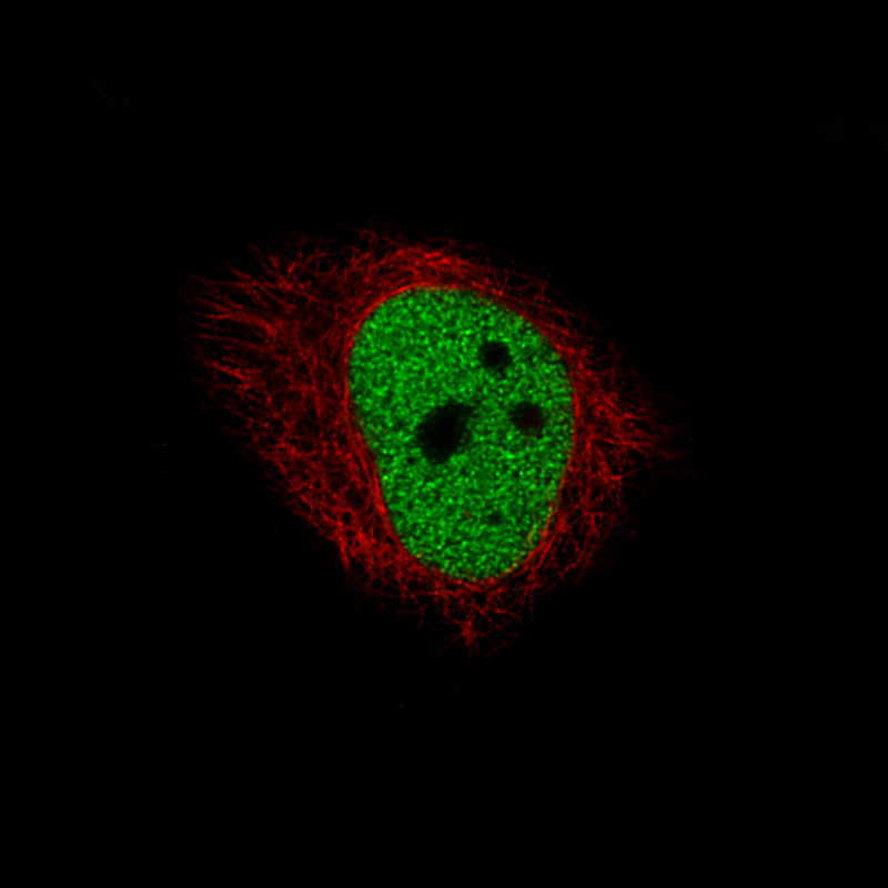
Immunofluorescence staining of U-251 cells using the Anti-HDAC1 monoclonal antibody, showing specific staining in the nucleoplasm in green. Microtubule- and nuclear probes are visualized in red and blue, respectively (where available). |
|
|
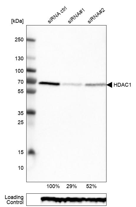
Western blot analysis in RT-4 cells transfected with control siRNA, target specific siRNA probe 1 and 2, using Anti-HDAC1 antibody. Remaining relative intensity is presented. Loading control: Anti-GAPDH. |
|
|
Immunohistochemical staining of human rectum shows strong nuclear positivity in glandular cells. |
|
|
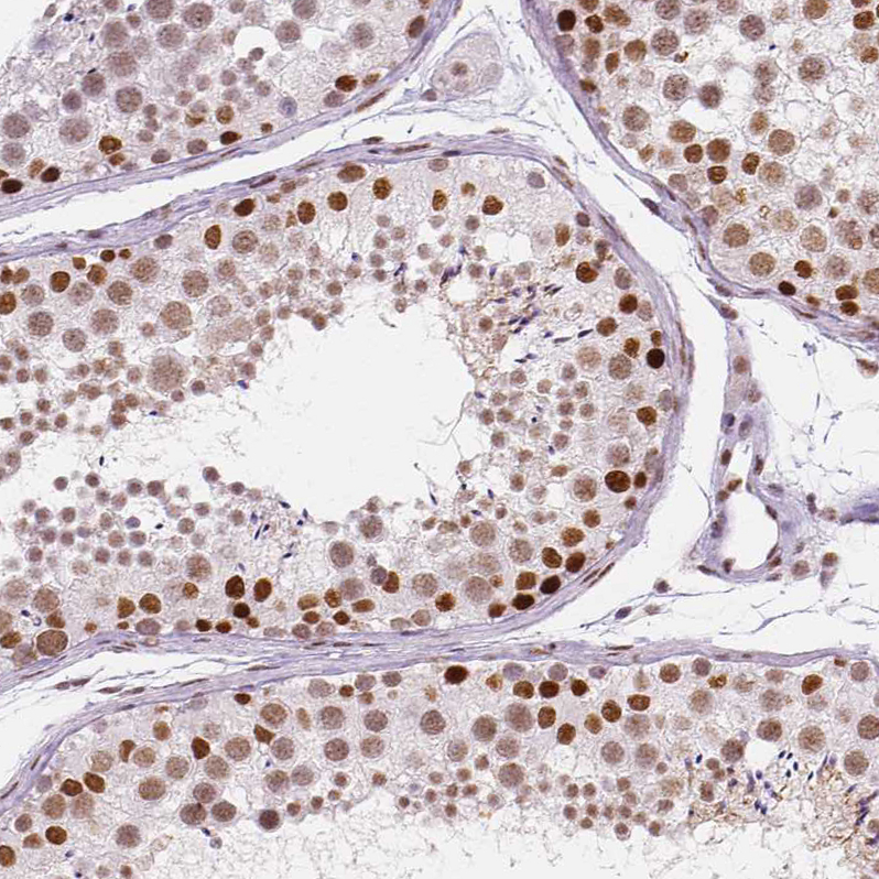
Immunohistochemical staining of human testis shows moderate to strong nuclear positivity in cells in seminiferous ducts. |
|
|
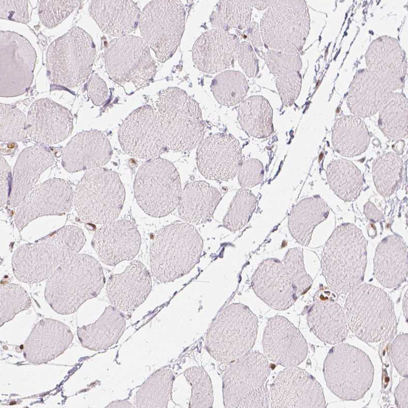
Immunohistochemical staining of human skeletal muscle shows no positivity in myocytes as expected. |
Product Guarantee and Expert Support
