
Anti-PDIA3 Recombinant Antibody, IgG1, Clone: [CL2444], Mouse, Monoclonal
Catalog Number:
ATA-AMAB90988R
- Images (12)
| Article Name: | Anti-PDIA3 Recombinant Antibody, IgG1, Clone: [CL2444], Mouse, Monoclonal |
| Biozol Catalog Number: | ATA-AMAB90988R |
| Supplier Catalog Number: | AMAb90988R |
| Alternative Catalog Number: | ATA-AMAB90988R-100, ATA-AMAB90988R-25 |
| Manufacturer: | Atlas Antibodies |
| Host: | Mouse |
| Category: | Antikörper |
| Application: | ICC, IHC, WB |
| Species Reactivity: | Human |
| Alternative Names: | ERp57, ERp60, ERp61, GRP57, GRP58, HsT17083, P58, PI-PLC |
| Recombinant Mouse Monoclonal Anti-PDIA3 Antibody against Human protein disulfide isomerase family A, member 3. Validated for Immunofluorescence, Immunohistochemistry and Western Blot |
|
|
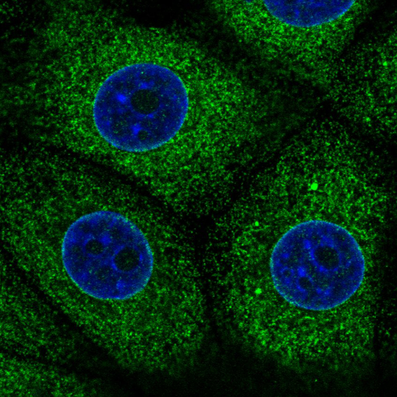
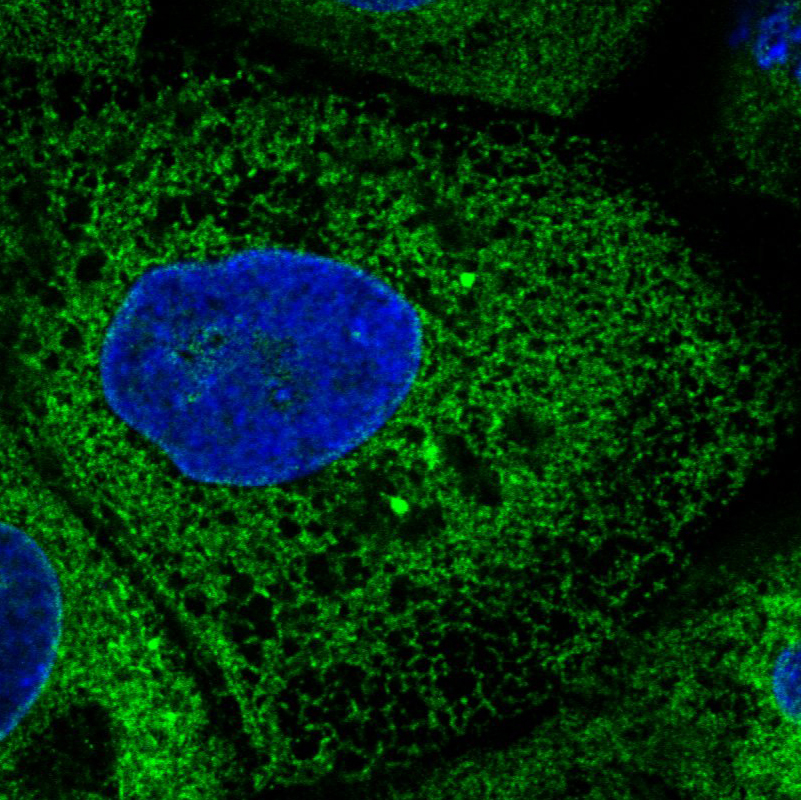
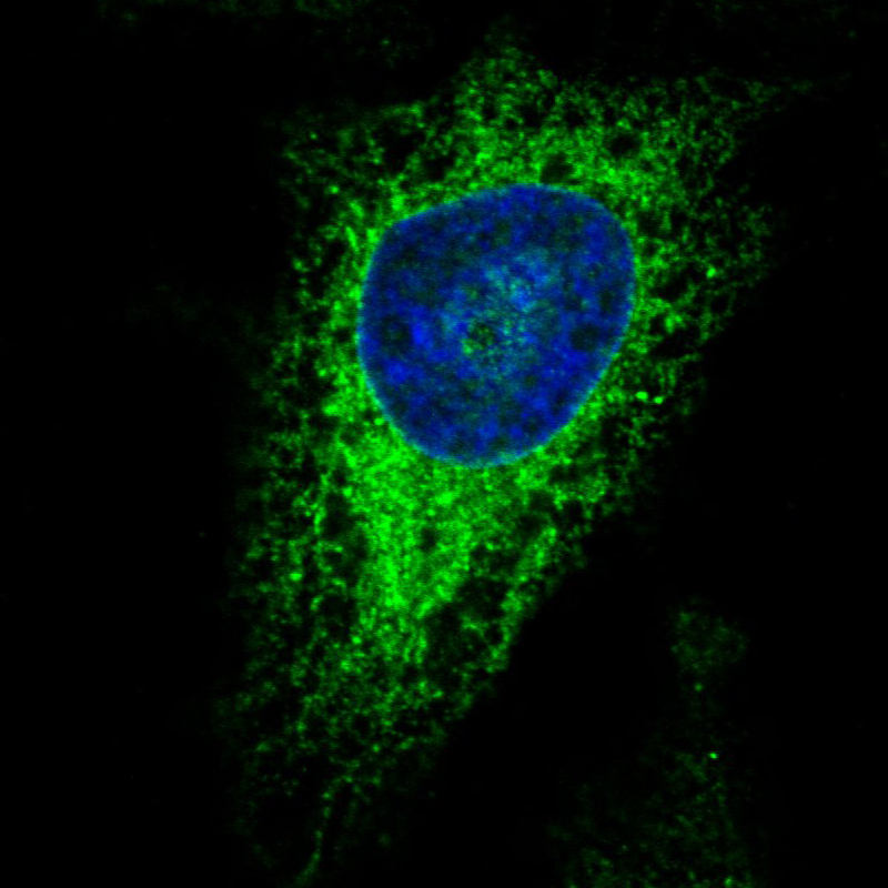
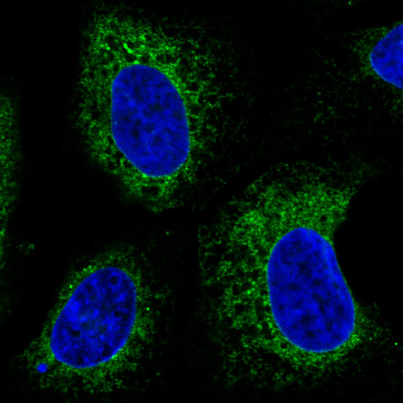
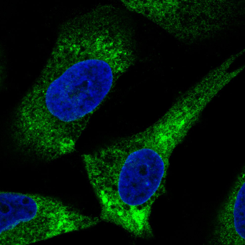
Immunofluorescence staining in MCF7 cell line with Anti-PDIA3 monoclonal antibody, showing specific staining of endoplasmic reticulum in green. Microtubule- and nuclear probes are visualized in red and blue respectively (where available). |
|
|
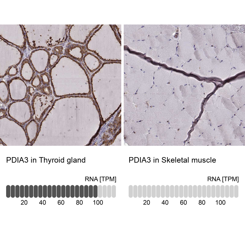
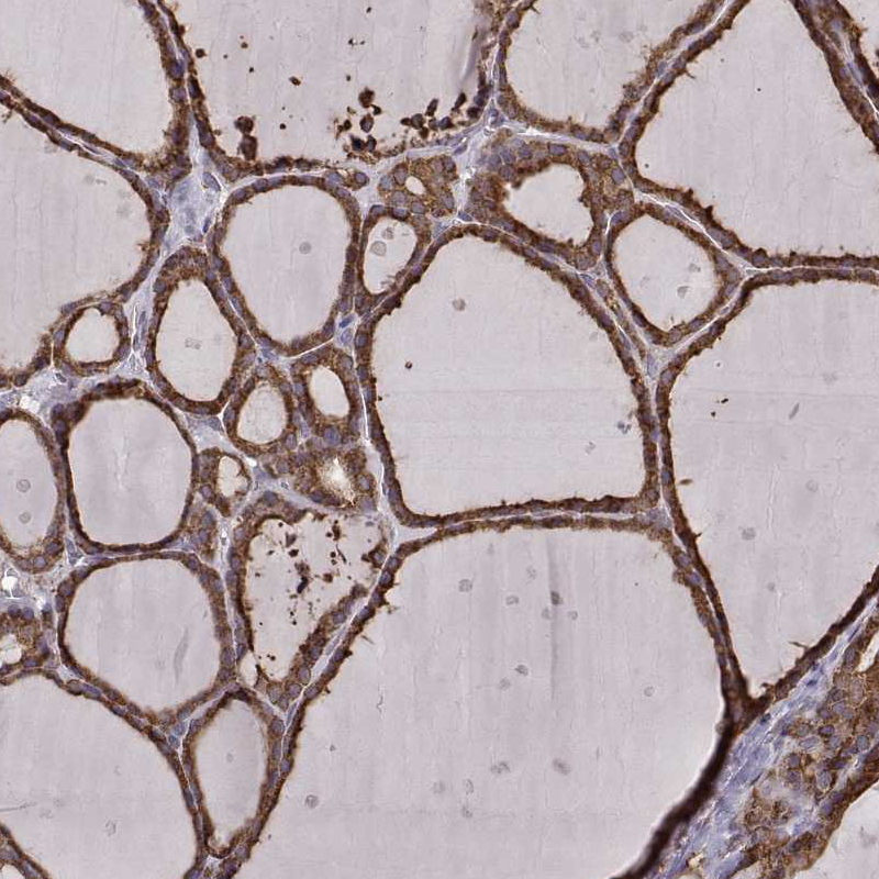
Immunohistochemistry analysis in human thyroid gland and skeletal muscle tissues using AMAb90988 antibody. Corresponding PDIA3 RNA-seq data are presented for the same tissues. |
|
|
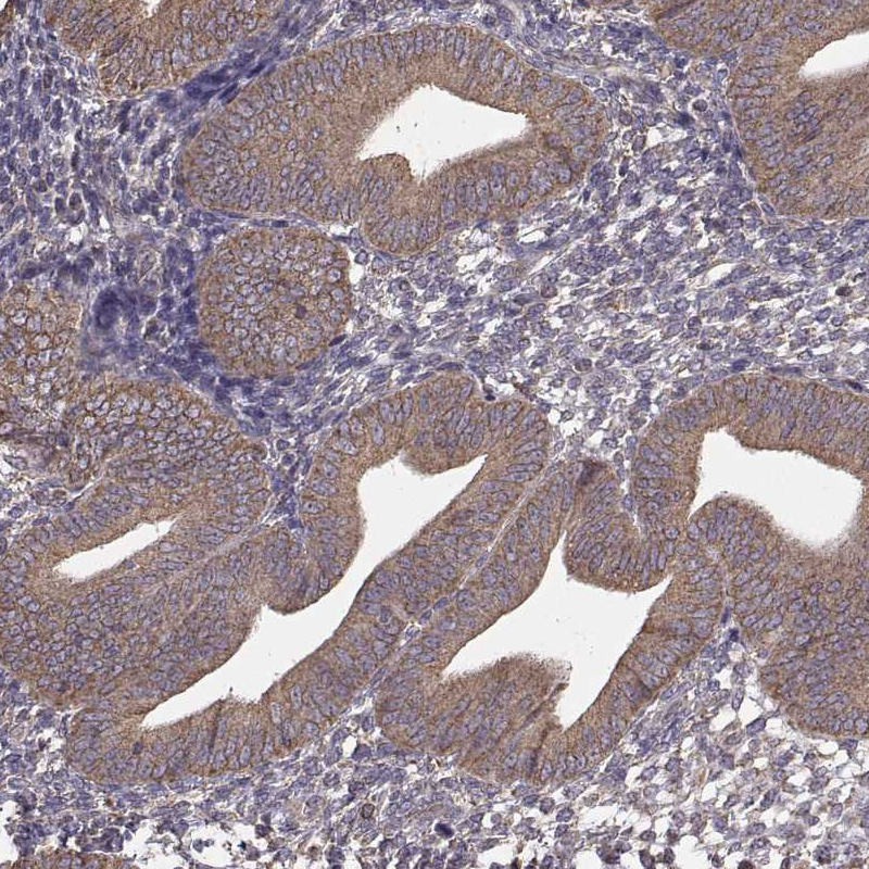
Immunohistochemical staining of human endometrium shows moderate cytoplasmic positivity in glandular cells. |
|
|
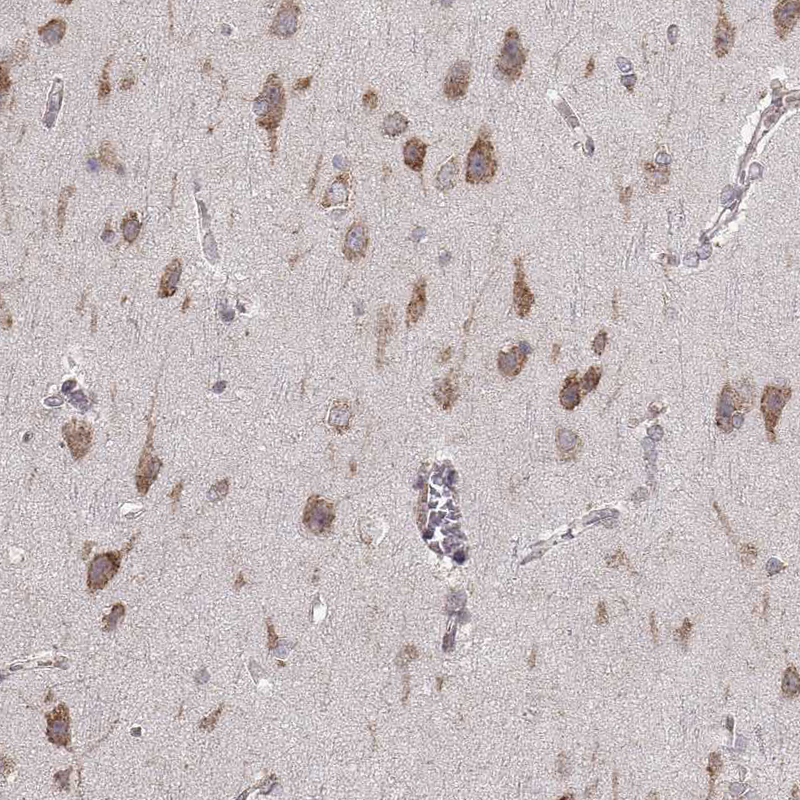
Immunohistochemical staining of human cerebral cortex shows moderate cytoplasmic positivity in neurons. |
|
|
Immunofluorescence staining in A431 cell line with Anti-PDIA3 monoclonal antibody, showing specific staining of endoplasmic reticulum in green. Microtubule- and nuclear probes are visualized in red and blue respectively (where available). |
|
|
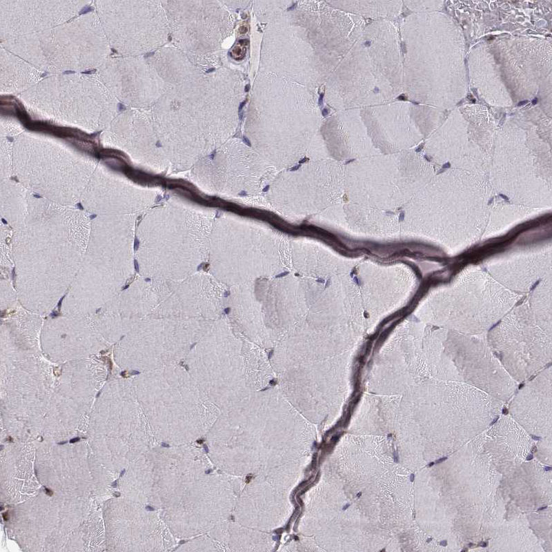
Immunohistochemical staining of human skeletal muscle shows no positivity in striated muscle fibers as expected. |
|
|
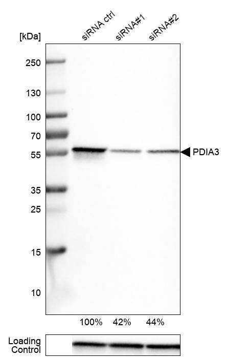
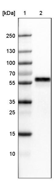
Western blot analysis in U-251MG cells transfected with control siRNA, target specific siRNA probe 1 and 2, using Anti-PDIA3 antibody. Remaining relative intensity is presented. Loading control: Anti-GAPDH. |
|
|
Immunofluorescence staining in HeLa cell line with Anti-PDIA3 monoclonal antibody, showing specific staining of endoplasmic reticulum in green. Microtubule- and nuclear probes are visualized in red and blue respectively (where available). |
|
|
Immunofluorescence staining in U2OS cell line with Anti-PDIA3 monoclonal antibody, showing specific staining of endoplasmic reticulum in green. Microtubule- and nuclear probes are visualized in red and blue respectively (where available). |
|
|
Lane 1: Marker [kDa] Lane 2:Human cell line U-251 MG |
|
|
Immunohistochemical staining of human thyroid gland shows moderate to strong cytoplasmic positivity in glandular cells. |
|
|
Immunofluorescence staining in U251 cell line with Anti-PDIA3 monoclonal antibody, showing specific staining of endoplasmic reticulum in green. Microtubule- and nuclear probes are visualized in red and blue respectively (where available). |
Product Guarantee and Expert Support
