
Anti-MBP Recombinant Antibody, IgG1, Clone: [CL2829], Mouse, Monoclonal
Catalog Number:
ATA-AMAB91064R
- Images (12)

| Article Name: | Anti-MBP Recombinant Antibody, IgG1, Clone: [CL2829], Mouse, Monoclonal |
| Biozol Catalog Number: | ATA-AMAB91064R |
| Supplier Catalog Number: | AMAb91064R |
| Alternative Catalog Number: | ATA-AMAB91064R-100, ATA-AMAB91064R-25 |
| Manufacturer: | Atlas Antibodies |
| Host: | Mouse |
| Category: | Antikörper |
| Application: | IHC, WB |
| Species Reactivity: | Human, Mouse, Rat |
| Recombinant Mouse Monoclonal Anti-MBP Antibody against Human myelin basic protein. Validated for Immunohistochemistry and Western Blot |
|
|
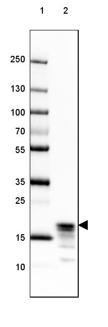
Lane 1: Marker [kDa] 250, 130, 100, 70, 55, 35, 25, 15, 10 Lane 2: Human Cerebral Cortex tissue |
|
|
Immunofluorescence staining of mouse hippocampus shows strong positivity in myelinated neural fibers. |
|
|
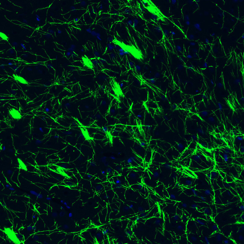
Immunofluorescence staining of mouse cerebellum shows strong positivity in myelinated neural fibers. |
|
|
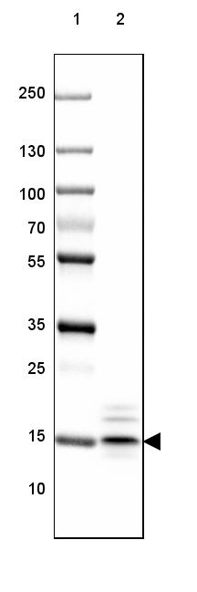
Lane 1: Marker [kDa] 250, 130, 100, 70, 55, 35, 25, 15, 10 Lane 2: Mouse Cerebral Cortex tissue |
|
|
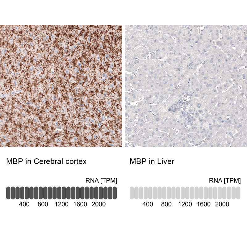
Immunohistochemistry analysis in human cerebral cortex and liver tissues using AMAb91064 antibody. Corresponding MBP RNA-seq data are presented for the same tissues. |
|
|
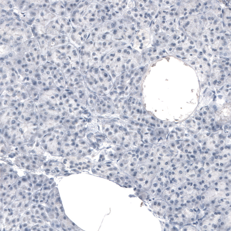
Immunohistochemical staining of human liver shows no positivity in hepatocytes as expected. |
|
|
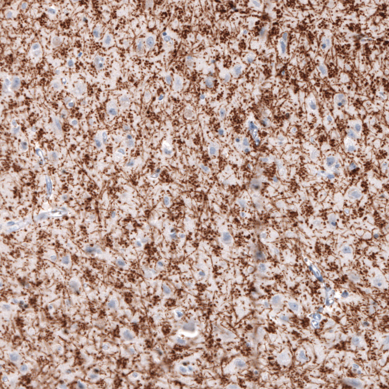
Immunohistochemical staining of rat cerebral cortex shows strong immunoreactivity in myelinated fibers. |
|
|
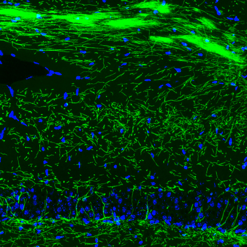
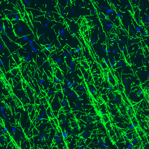
Immunofluorescence staining of rat basal forebrain shows strong positivity in myelinated neural fibers. |
|
|
Immunohistochemical staining of human pancreas shows no positivity in exocrine glandular cells as expected. |
|
|
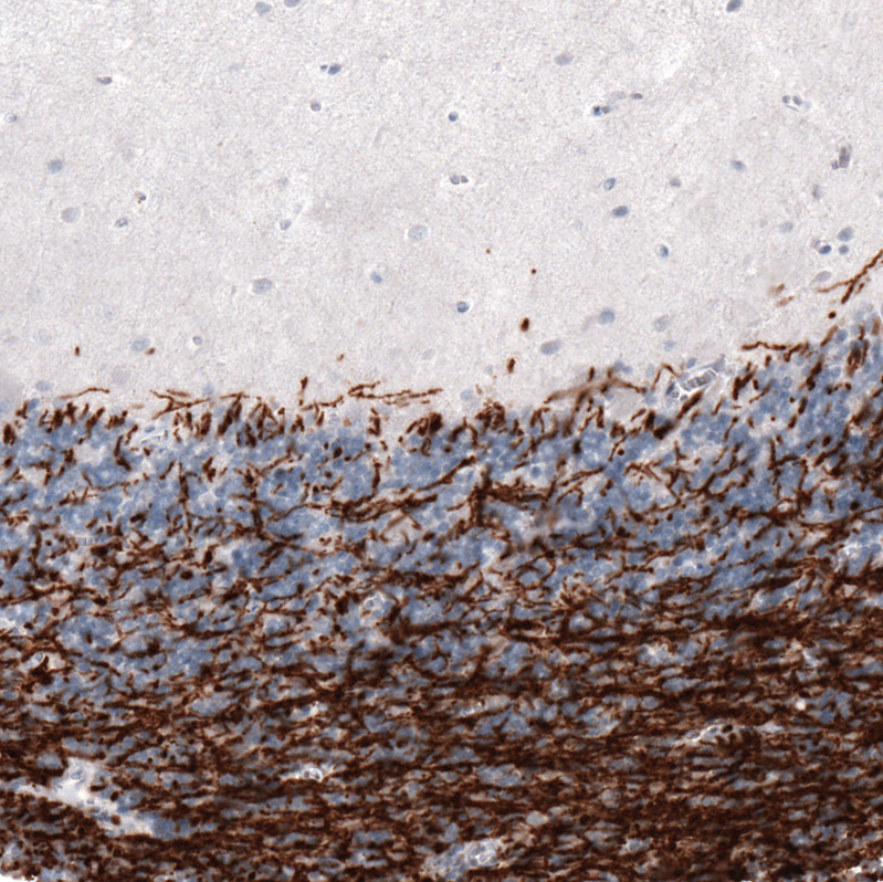
Immunohistochemical staining of human cerebral cortex shows strong positivity in myelinated neural fibers. |
|
|
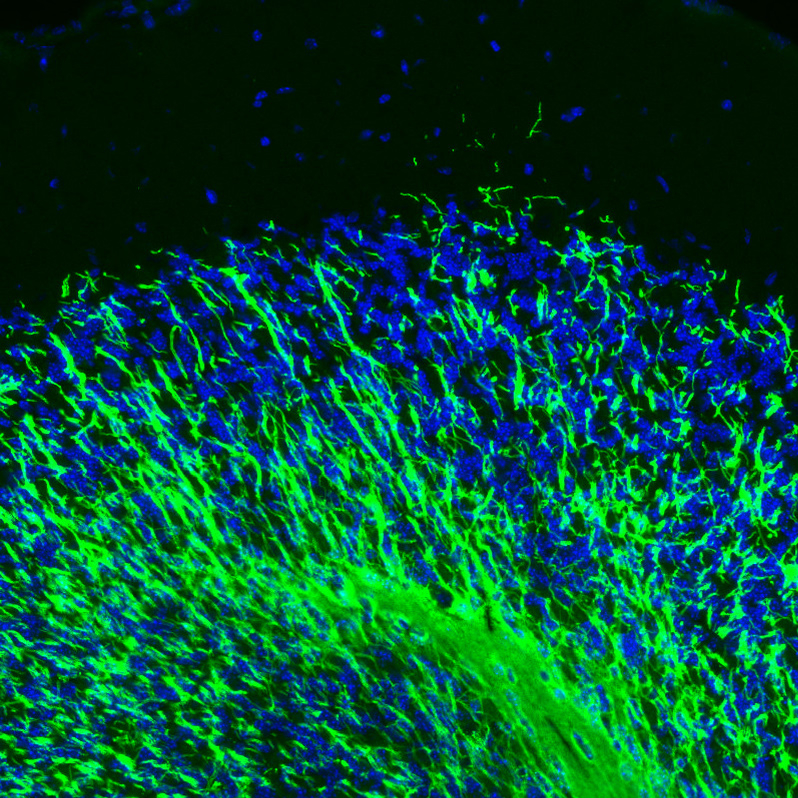
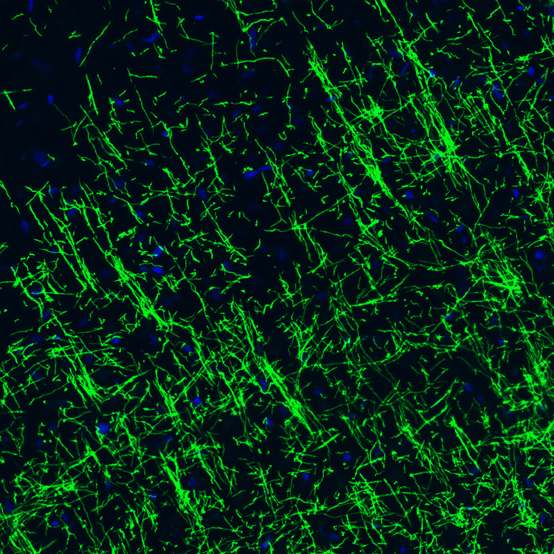
Immunofluorescence staining of rat cerebral cortex shows strong positivity in myelinated neural fibers. |
|
|
Immunohistochemical staining of human cerebellum shows strong positivity in myelinated neural fiebrs. |
Product Guarantee and Expert Support
