
Anti-TUBB3 Recombinant Antibody, IgG1, Clone: [CL5813], Mouse, Monoclonal
Catalog Number:
ATA-AMAB91394R
- Images (11)
| Article Name: | Anti-TUBB3 Recombinant Antibody, IgG1, Clone: [CL5813], Mouse, Monoclonal |
| Biozol Catalog Number: | ATA-AMAB91394R |
| Supplier Catalog Number: | AMAb91394R |
| Alternative Catalog Number: | ATA-AMAB91394R-100, ATA-AMAB91394R-25 |
| Manufacturer: | Atlas Antibodies |
| Host: | Mouse |
| Category: | Antikörper |
| Application: | ICC, IHC, WB |
| Species Reactivity: | Human, Mouse, Rat |
| Alternative Names: | beta-4, CFEOM3, CFEOM3A, FEOM3 |
| Recombinant Mouse Monoclonal Anti-TUBB3 Antibody against Human Tubulin beta 3 class iii. Validated for Immunofluorescence, Immunohistochemistry and Western Blot |
|
|
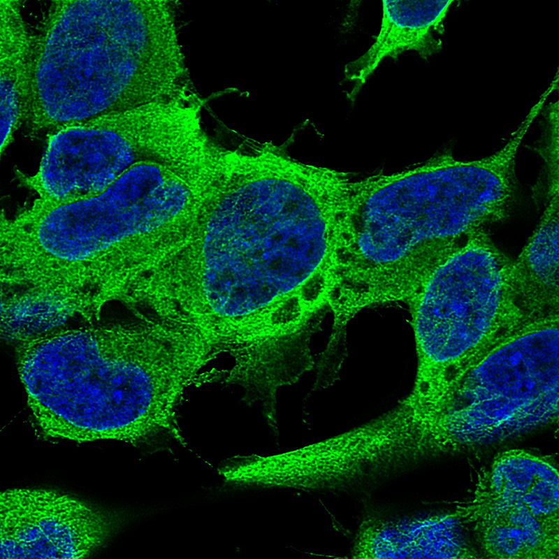
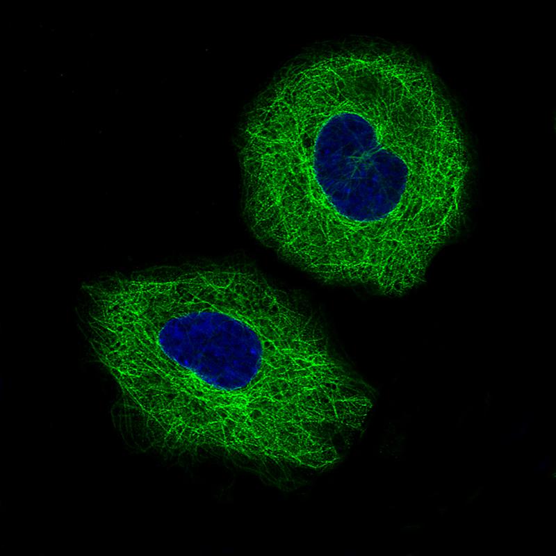
Immunofluorescence staining of SH-SY5Y cells using the Anti-TUBB3 monoclonal antibody, showing specific staining on microtubules in green. Nuclear probe is visualized in blue. |
|
|
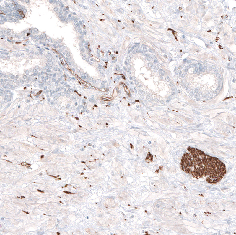
Immunohistochemical staining of human prostate shows strong positivity in peripheral nerves. |
|
|
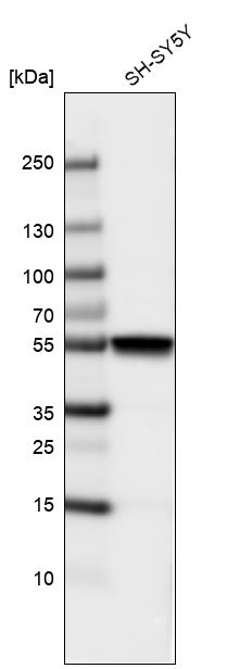
Western blot analysis in human cell line SH-SY5Y. |
|
|
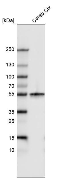
Western blot analysis in human cerebral cortex tissue. |
|
|
Immunohistochemical staining of human cerebellum shows strong positivity in neurons and neuropil. |
|
|
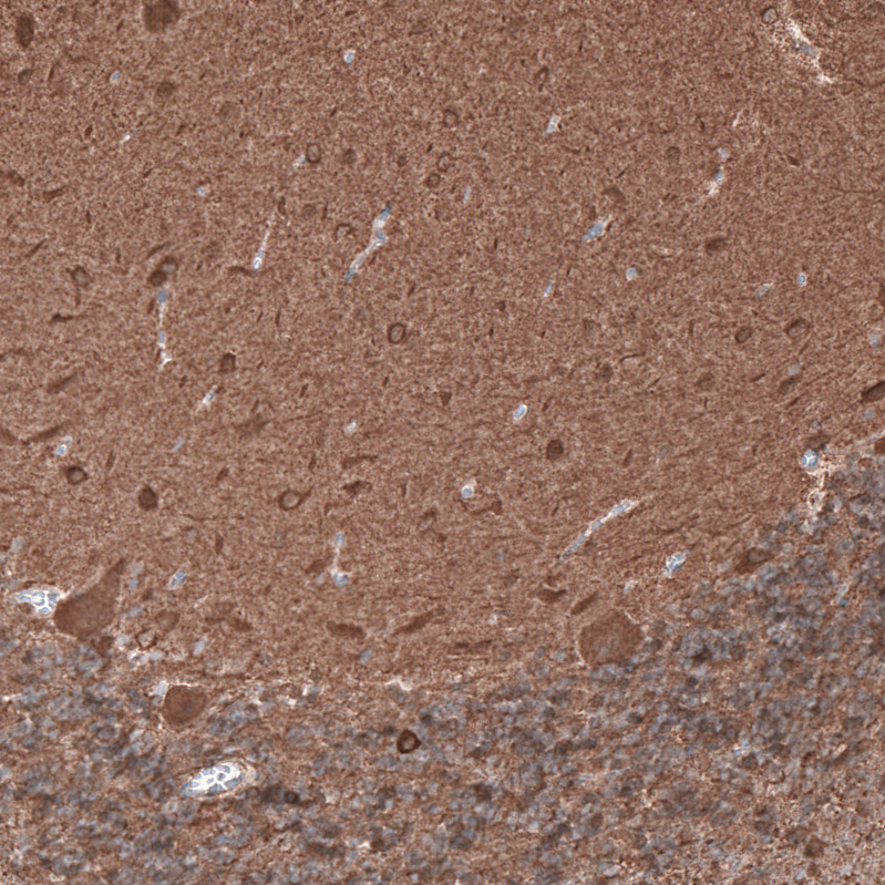
Immunohistochemical staining of rat brain shows strong cytoplasmic positivity in neurons. |
|
|
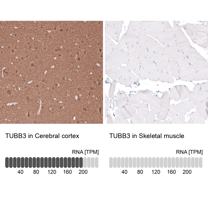
Immunohistochemistry analysis in human cerebral cortex and skeletal muscle tissues using AMAb91394 antibody. Corresponding TUBB3 RNA-seq data are presented for the same tissues. |
|
|
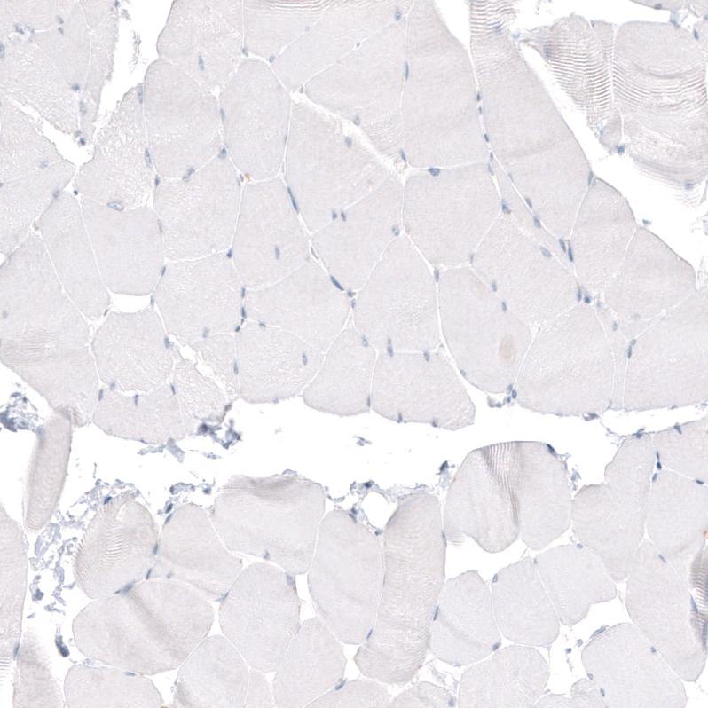
Immunohistochemical staining of human skeletal muscle shows no positivity in myocytes as expected. |
|
|
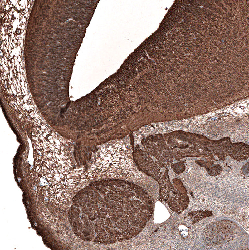
Immunohistochemical staining of mouse embryo E11 shows strong positivity in the developing central and peripheral nervous system. |
|
|
Immunofluorescence staining of A549 cells using the Anti-TUBB3 monoclonal antibody, showing specific staining on microtubules in green. Microtubule- and nuclear probes are visualized in red and blue, respectively (where available). |
|
|
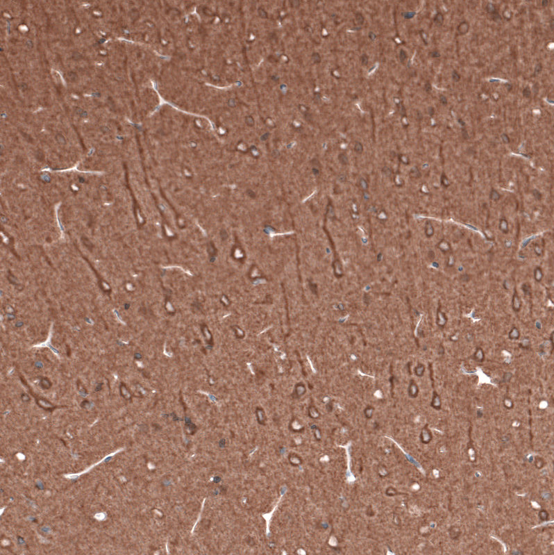
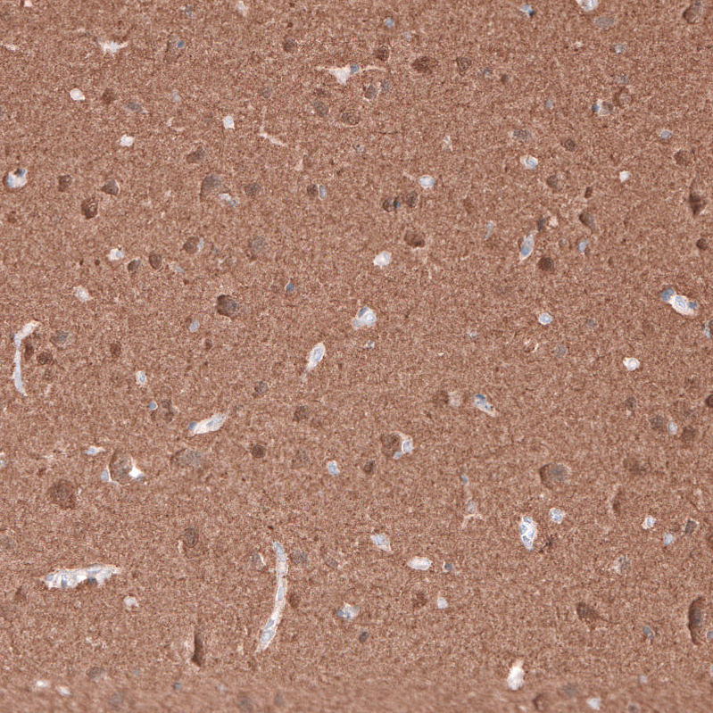
Immunohistochemical staining of human cerebral cortex shows strong positivity in neurons and neuropil. |
Product Guarantee and Expert Support
