
Anti-CD44 Antibody Picoband, Rabbit, Polyclonal
Catalog Number:
BOB-A00052-CARRIER-FREE
- Images (37)


| Article Name: | Anti-CD44 Antibody Picoband, Rabbit, Polyclonal |
| Biozol Catalog Number: | BOB-A00052-CARRIER-FREE |
| Supplier Catalog Number: | A00052-carrier-free |
| Alternative Catalog Number: | BOB-A00052-CARRIER-FREE-100UG |
| Manufacturer: | Boster Bio |
| Host: | Rabbit |
| Category: | Antikörper |
| Application: | ELISA, FC, ICC, IF, IHC, IHC-Fr, WB |
| Species Reactivity: | Human, Mouse, Rat |
| Immunogen: | E. coli-derived human CD44 recombinant protein (Position: Q21-H259). |
| Alternative Names: | CD44, CD44 antigen, CDW44, CSPG8, ECMR III, Epican, HCELL, Heparan sulfate proteoglycan, Hermes antigen, HUTCH I, Hyaluronate receptor, IN, MC56, MDU2, MDU3, MIC4, MUTCH I, PGP 1, PGP I, Pgp1, Phagocytic glycoprotein 1, Phagocytic glycoprotein I |
| Boster Bio Anti-CD44 Antibody Picoband catalog A00052. Tested in ELISA, Flow Cytometry, IF, IHC, IHC-F, ICC, WB applications. This antibody reacts with Human, Mouse, Rat. The brand Picoband indicates this is a premium antibody that guarantees superior quality, high affinity, and strong signals with minimal background in Western blot applications. Only our best-performing antibodies are designated as Picoband, ensuring unmatched performance. |
| Clonality: | Polyclonal |
| Concentration: | Adding 0.2 ml of distilled water will yield a concentration of 500 µg/ml. |
| Molecular Weight: | Observed Molecular Weight: 82 kDa. Calculated Molecular Weight: 25283 MW |
| NCBI: | 960 |
| UniProt: | P16070 |
| Buffer: | Each vial contains 4mg Trehalose, 0.9mg NaCl, 0.2mg Na2HPO4, 0.05mg NaN3. |
| Purity: | Immunogen affinity purified. |
| Form: | Lyophilized |
| Target: | CD44 antigen |
| Application Dilute: | Western blot, 0.1-0.5µg/ml Immunohistochemistry (Paraffin-embedded Section), 0.5-1µg/ml Immunohistochemistry (Frozen Section), 0.5-1µg/ml Immunocytochemistry, 0.5-1µg/ml Immunofluorescence, 2µg/ml Flow Cytometry (Fixed), 1-3µg/1x106 cells ELISA, 0.1-0.5µg |
|
|
|
|
|
|
|
|
|
|
|
|
|
|
|
|
|
|
|
|
|
|
|
|
|
|
|
|
|
|
|
|
|
|
|
|
|
|
|
|
|
|
|
|
|
|
|
|
|
|
|
|
|
|
|
|
|
|
|
|
|
|
|
|
|
|
|
|
|
|
|
|
|
|
|
|
|
|
|
|
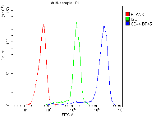
Flow Cytometry analysis of Jurkat cells using anti-CD44 antibody (A00052). Overlay histogram showing Jurkat cells stained with A00052 (Blue line). To facilitate intracellular staining, cells were fixed with 4% paraformaldehyde and permeabilized with permeabilization buffer. The cells were blocked with 10% normal goat serum. And then incubated with rabbit anti-CD44 Antibody (A00052&44,1µg/1x106 cells) for 30 min at 20C. DyLight488 conjugated goat anti-rabbit IgG (BA1127&44, 5-10µg/1x106 cells) was used as secondary antibody for 30 minutes at 20C. Isotype control antibody (Green line) was rabbit IgG (1µg/1x106) used under the same conditions. Unlabelled sample without incubation with primary antibody and secondary antibody (Red line) was used as a blank... |
|
|
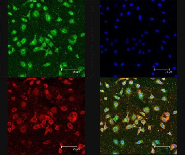
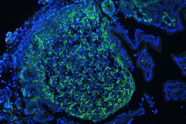
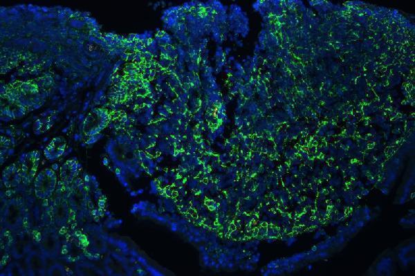
IF analysis of CD44 using anti-CD44 antibody (A00052) CD44 was detected in paraffin-embedded section of mouse lymphaden tissues. Heat mediated antigen retrieval was performed in citrate buffer (pH6&44, epitope retrieval solution ) for 20 mins. The tissue section was blocked with 10% goat serum. The tissue section was then incubated with 1µg/mL rabbit anti-CD44 Antibody (A00052) overnight at 4C. DyLight488 Conjugated Goat Anti-Rabbit IgG (BA1127) was used as secondary antibody at 1:100 dilution and incubated for 30 minutes at 37C. The section was counterstained with DAPI. Visualize using a fluorescence microscope and filter sets appropriate for the label use |
|
|
|
|
|
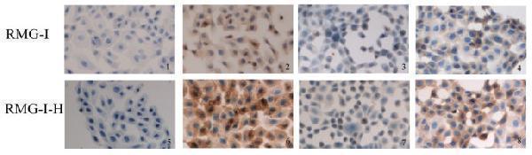
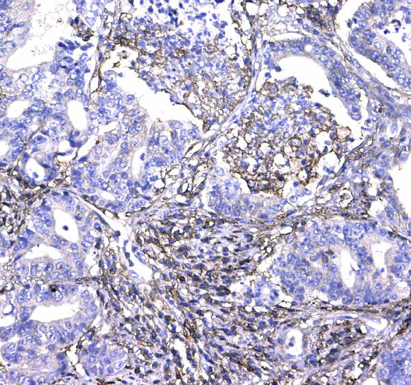
IHC analysis of CD44 using anti-CD44 antibody (A00052). CD44 was detected in paraffin-embedded section of human colon cancer tissue. Heat mediated antigen retrieval was performed in citrate buffer (pH6&44, epitope retrieval solution) for 20 mins. The tissue section was blocked with 10% goat serum. The tissue section was then incubated with 1µg/ml rabbit anti-CD44 Antibody (A00052) overnight at 4C. Biotinylated goat anti-rabbit IgG was used as secondary antibody and incubated for 30 minutes at 37C. The tissue section was developed using Strepavidin-Biotin-Complex (SABC)(Catalog SA1022) with DAB as the chromogen. |
|
|
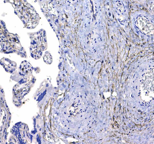
IHC analysis of CD44 using anti-CD44 antibody (A00052). CD44 was detected in paraffin-embedded section of human placenta tissue . Heat mediated antigen retrieval was performed in citrate buffer (pH6&44, epitope retrieval solution) for 20 mins. The tissue section was blocked with 10% goat serum. The tissue section was then incubated with 1µg/ml rabbit anti-CD44 Antibody (A00052) overnight at 4C. Biotinylated goat anti-rabbit IgG was used as secondary antibody and incubated for 30 minutes at 37C. The tissue section was developed using Strepavidin-Biotin-Complex (SABC)(Catalog SA1022) with DAB as the chromogen. |
|
|
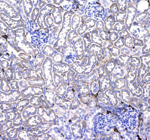
IHC analysis of CD44 using anti-CD44 antibody (A00052). CD44 was detected in paraffin-embedded section of mouse kidney tissue . Heat mediated antigen retrieval was performed in citrate buffer (pH6&44, epitope retrieval solution) for 20 mins. The tissue section was blocked with 10% goat serum. The tissue section was then incubated with 1µg/ml rabbit anti-CD44 Antibody (A00052) overnight at 4C. Biotinylated goat anti-rabbit IgG was used as secondary antibody and incubated for 30 minutes at 37C. The tissue section was developed using Strepavidin-Biotin-Complex (SABC)(Catalog SA1022) with DAB as the chromogen. |
|
|
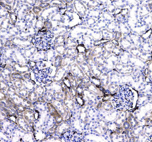
IHC analysis of CD44 using anti-CD44 antibody (A00052). CD44 was detected in paraffin-embedded section of rat kidney tissue . Heat mediated antigen retrieval was performed in citrate buffer (pH6&44, epitope retrieval solution) for 20 mins. The tissue section was blocked with 10% goat serum. The tissue section was then incubated with 1µg/ml rabbit anti-CD44 Antibody (A00052) overnight at 4C. Biotinylated goat anti-rabbit IgG was used as secondary antibody and incubated for 30 minutes at 37C. The tissue section was developed using Strepavidin-Biotin-Complex (SABC)(Catalog SA1022) with DAB as the chromogen. |
|
|
|
|
|
|
|
|
|
|
|
Product Guarantee and Expert Support
