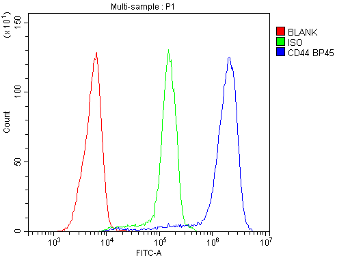
Flow Cytometry analysis of Jurkat cells using anti-CD44 antibody (A00052). Overlay histogram showing Jurkat cells stained with A00052 (Blue line). To facilitate intracellular staining, cells were fixed with 4% paraformaldehyde and permeabilized with permeabilization buffer. The cells were blocked with 10% normal goat serum. And then incubated with rabbit anti-CD44 Antibody (A00052&44,1µg/1x106 cells) for 30 min at 20C. DyLight488 conjugated goat anti-rabbit IgG (BA1127&44, 5-10µg/1x106 cells) was used as secondary antibody for 30 minutes at 20C. Isotype control antibody (Green line) was rabbit IgG (1µg/1x106) used under the same conditions. Unlabelled sample without incubation with primary antibody and secondary antibody (Red line) was used as a blank control.
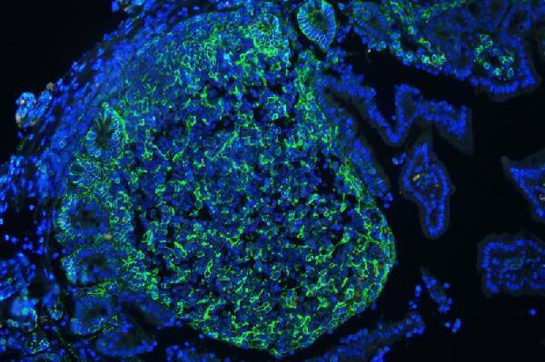
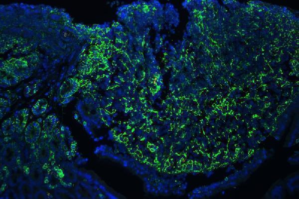
IF analysis of CD44 using anti-CD44 antibody (A00052) CD44 was detected in paraffin-embedded section of mouse lymphaden tissues. Heat mediated antigen retrieval was performed in citrate buffer (pH6&44, epitope retrieval solution ) for 20 mins. The tissue section was blocked with 10% goat serum. The tissue section was then incubated with 1µg/mL rabbit anti-CD44 Antibody (A00052) overnight at 4C. DyLight488 Conjugated Goat Anti-Rabbit IgG (BA1127) was used as secondary antibody at 1:100 dilution and incubated for 30 minutes at 37C. The section was counterstained with DAPI. Visualize using a fluorescence microscope and filter sets appropriate
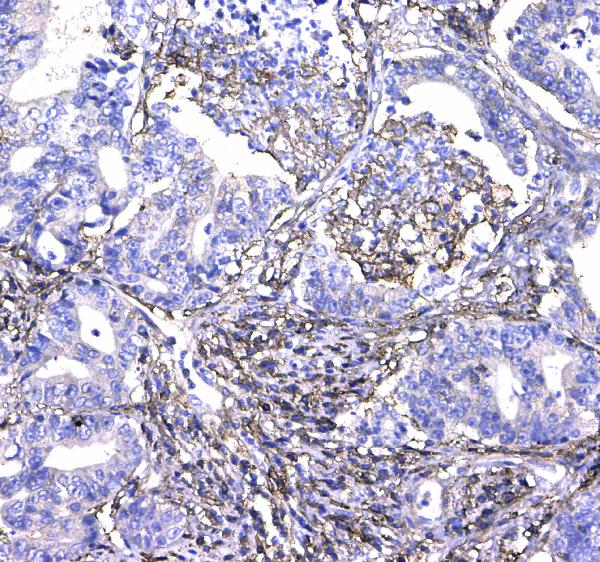
IHC analysis of CD44 using anti-CD44 antibody (A00052).CD44 was detected in paraffin-embedded section of human colon cancer tissue. Heat mediated antigen retrieval was performed in citrate buffer (pH6&44, epitope retrieval solution) for 20 mins. The tissue section was blocked with 10% goat serum. The tissue section was then incubated with 1µg/ml rabbit anti-CD44 Antibody (A00052) overnight at 4C. Biotinylated goat anti-rabbit IgG was used as secondary antibody and incubated for 30 minutes at 37C. The tissue section was developed using Strepavidin-Biotin-Complex (SABC)(Catalog SA1022) with DAB as the chromogen.
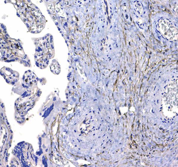
IHC analysis of CD44 using anti-CD44 antibody (A00052).CD44 was detected in paraffin-embedded section of human placenta tissue . Heat mediated antigen retrieval was performed in citrate buffer (pH6&44, epitope retrieval solution) for 20 mins. The tissue section was blocked with 10% goat serum. The tissue section was then incubated with 1µg/ml rabbit anti-CD44 Antibody (A00052) overnight at 4C. Biotinylated goat anti-rabbit IgG was used as secondary antibody and incubated for 30 minutes at 37C. The tissue section was developed using Strepavidin-Biotin-Complex (SABC)(Catalog SA1022) with DAB as the chromogen.
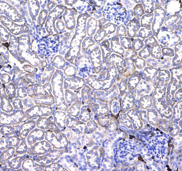
IHC analysis of CD44 using anti-CD44 antibody (A00052).CD44 was detected in paraffin-embedded section of mouse kidney tissue . Heat mediated antigen retrieval was performed in citrate buffer (pH6&44, epitope retrieval solution) for 20 mins. The tissue section was blocked with 10% goat serum. The tissue section was then incubated with 1µg/ml rabbit anti-CD44 Antibody (A00052) overnight at 4C. Biotinylated goat anti-rabbit IgG was used as secondary antibody and incubated for 30 minutes at 37C. The tissue section was developed using Strepavidin-Biotin-Complex (SABC)(Catalog SA1022) with DAB as the chromogen.
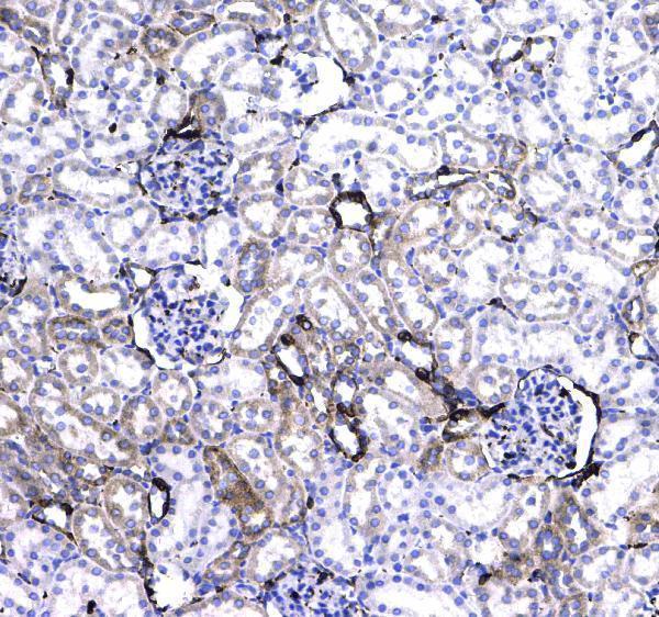
IHC analysis of CD44 using anti-CD44 antibody (A00052).CD44 was detected in paraffin-embedded section of rat kidney tissue . Heat mediated antigen retrieval was performed in citrate buffer (pH6&44, epitope retrieval solution) for 20 mins. The tissue section was blocked with 10% goat serum. The tissue section was then incubated with 1µg/ml rabbit anti-CD44 Antibody (A00052) overnight at 4C. Biotinylated goat anti-rabbit IgG was used as secondary antibody and incubated for 30 minutes at 37C. The tissue section was developed using Strepavidin-Biotin-Complex (SABC)(Catalog SA1022) with DAB as the chromogen.
* VAT and and shipping costs not included. Errors and price changes excepted