
Anti-UBE2D1/2/3/4 Antibody Picoband, Rabbit, Polyclonal
Catalog Number:
BOB-A04728-1
- Images (9)

| Article Name: | Anti-UBE2D1/2/3/4 Antibody Picoband, Rabbit, Polyclonal |
| Biozol Catalog Number: | BOB-A04728-1 |
| Supplier Catalog Number: | A04728-1 |
| Alternative Catalog Number: | BOB-A04728-1-100UG |
| Manufacturer: | Boster Bio |
| Host: | Rabbit |
| Category: | Antikörper |
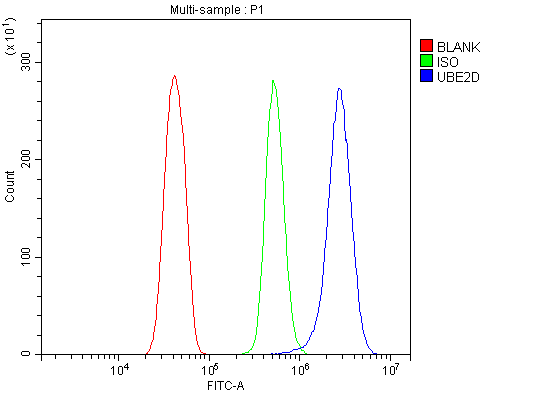
| Application: | ELISA, FC, ICC, IF, IHC, WB |
| Species Reactivity: | Human, Mouse, Rat |
| Immunogen: | E.coli-derived human UBE2D1/2/3/4 recombinant protein (Position: D116-M147). |
| Alternative Names: | E2(17)KB1, SFT, Stimulator of Fe transport, UBC4/5, UBC4/5 homolog, UBC5A, UBCH5, UBCH5A, UBE2D, UBE2D1, UBE2D1/2/3/4, UBE2D2, UBE2D3, UBE2D4, Ubiquitin carrier protein D1, Ubiquitin protein ligase D1, E2(17)KB2, PUBC1, UBC4, UBC5B, UBCH4, UBCH5B, Ubiquitin carrier protein D2, Ubiquitin protein ligase D2, E2(17)KB3, UBC5C, UBCH5C, Ubiquitin carrier protein D3, Ubiquitin protein ligase D3, HBUCE1, UBCH5D, Ubiquitin carrier protein D4, Ubiquitin protein ligase D4 |
| Boster Bio Anti-UBE2D1/2/3/4 Antibody Picoband catalog A04728-1. Tested in ELISA, Flow Cytometry, IF, IHC, ICC, WB applications. This antibody reacts with Human, Mouse, Rat. The brand Picoband indicates this is a premium antibody that guarantees superior quality, high affinity, and strong signals with minimal background in Western blot applications. Only our best-performing antibodies are designated as Picoband, ensuring unmatched performance. |
| Clonality: | Polyclonal |
| Concentration: | Adding 0.2 ml of distilled water will yield a concentration of 500 µg/ml. |
| Molecular Weight: | Observed Molecular Weight: 17 kDa. Calculated Molecular Weight: 32193 MW |
| NCBI: | 7321 |
| UniProt: | P51668 |
| Buffer: | Each vial contains 4mg Trehalose, 0.9mg NaCl, 0.2mg Na2HPO4, 0.005mg NaN3. |
| Purity: | Immunogen affinity purified. |
| Form: | Lyophilized |
| Target: | Ly6-A antigen |
| Application Dilute: | Western blot, 0.25-0.5µg/ml, Human, Rat Immunohistochemistry (Paraffin-embedded Section), 2-5µg/ml, Human, Mouse, Rat Immunocytochemistry/Immunofluorescence, 5µg/ml, Human Flow Cytometry (Fixed), 1-3µg/1x106 cells, Human, Mouse, Rat ELISA, 0.1-0.5µg/ml, - |
|
|
|
|
|
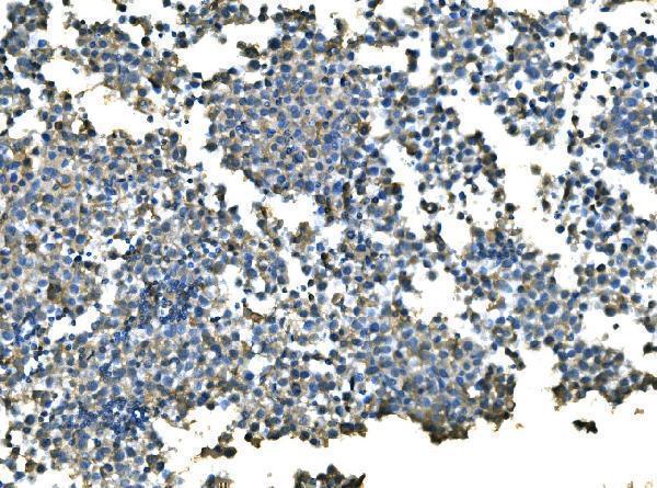
IHC analysis of UBE2D1/2/3/4 using anti-UBE2D1/2/3/4 antibody (A04728-1). UBE2D1/2/3/4 was detected in paraffin-embedded section of mouse lung lymph node tissue. Heat mediated antigen retrieval was performed in EDTA buffer (pH8.0, epitope retrieval solution). The tissue section was blocked with 10% goat serum. The tissue section was then incubated with 2µg/ml rabbit anti-UBE2D1/2/3/4 Antibody (A04728-1) overnight at 4C. Biotinylated goat anti-rabbit IgG was used as secondary antibody and incubated for 30 minutes at 37C. The tissue section was developed using Strepavidin-Biotin-Complex (SABC) (Catalog SA1022) with DAB as the chromogen. |
|
|
IHC analysis of UBE2D1/2/3/4 using anti-UBE2D1/2/3/4 antibody (A04728-1). UBE2D1/2/3/4 was detected in paraffin-embedded section of human testicular cancer tissue. Heat mediated antigen retrieval was performed in EDTA buffer (pH8.0, epitope retrieval solution). The tissue section was blocked with 10% goat serum. The tissue section was then incubated with 2µg/ml rabbit anti-UBE2D1/2/3/4 Antibody (A04728-1) overnight at 4C. Biotinylated goat anti-rabbit IgG was used as secondary antibody and incubated for 30 minutes at 37C. The tissue section was developed using Strepavidin-Biotin-Complex (SABC) (Catalog SA1022) with DAB as the chromogen. |
|
|
IHC analysis of UBE2D1/2/3/4 using anti-UBE2D1/2/3/4 antibody (A04728-1). UBE2D1/2/3/4 was detected in paraffin-embedded section of human bladder cancer tissue. Heat mediated antigen retrieval was performed in EDTA buffer (pH8.0, epitope retrieval solution). The tissue section was blocked with 10% goat serum. The tissue section was then incubated with 2µg/ml rabbit anti-UBE2D1/2/3/4 Antibody (A04728-1) overnight at 4C. Biotinylated goat anti-rabbit IgG was used as secondary antibody and incubated for 30 minutes at 37C. The tissue section was developed using Strepavidin-Biotin-Complex (SABC) (Catalog SA1022) with DAB as the chromogen. |
|
|
IHC analysis of UBE2D1/2/3/4 using anti-UBE2D1/2/3/4 antibody (A04728-1). UBE2D1/2/3/4 was detected in paraffin-embedded section of rat lung tissue. Heat mediated antigen retrieval was performed in EDTA buffer (pH8.0, epitope retrieval solution). The tissue section was blocked with 10% goat serum. The tissue section was then incubated with 2µg/ml rabbit anti-UBE2D1/2/3/4 Antibody (A04728-1) overnight at 4C. Biotinylated goat anti-rabbit IgG was used as secondary antibody and incubated for 30 minutes at 37C. The tissue section was developed using Strepavidin-Biotin-Complex (SABC) (Catalog SA1022) with DAB as the chromogen. |
|
|
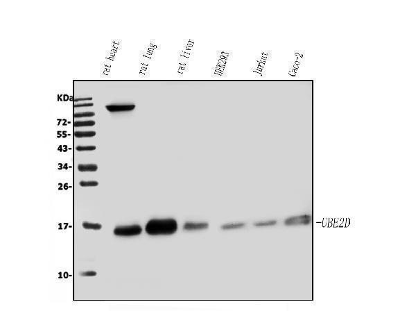
Western blot analysis of UBE2D1/2/3/4 using anti-UBE2D1/2/3/4 antibody (A04728-1). Electrophoresis was performed on a 5-20% SDS-PAGE gel at 70V (Stacking gel) / 90V (Resolving gel) for 2-3 hours. The sample well of each lane was loaded with 50ug of sample under reducing conditions. Lane 1: rat heart tissue lysates, Lane 2: rat lung tissue lysates, Lane 3: rat liver tissue lysates, Lane 4: human HEK293 whole cell lysates, Lane 5: human Jurkat whole cell lysates, Lane 6: human CACO-2 whole cell lysates. After Electrophoresis, proteins were transferred to a Nitrocellulose membrane at 150mA for 50-90 minutes. Blocked the membrane with 5% Non-fat Milk/ TBS for 1.5 hour at RT. The membrane was incubated with rabbit anti-UBE2D1/2/3/4 antigen affinity purified polyclonal antibody (Catalog A04728-1) at 0.5 µg/mL overnight at 4C, then washed with TBS-0.1%Tween 3 times with 5 minutes each and probed with a goat anti-rabbit IgG-HRP secondary antibody at a dilution of 1:5000 for 1.5 hour at RT. The signal is developed using an Enhanced Chemiluminescent detection (ECL) kit (Catalog EK1002) with Tanon 5200 system. A specific band was detected for UBE2D1/2/3/4 at approximately 17KD. The expected band size for UBE2D1/2/3/4 is at 17KD. |
|
|
|
|
|
|
|
|
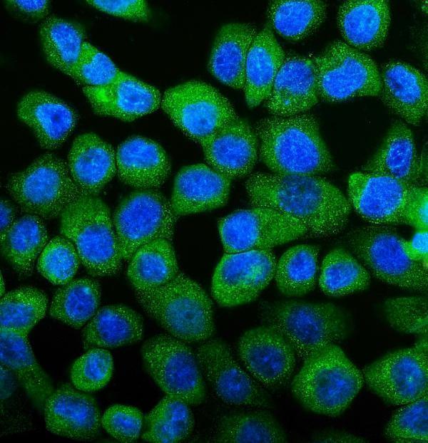
IF analysis of UBE2D1/2/3/4 using anti-UBE2D1/2/3/4 antibody (A04728-1). UBE2D1/2/3/4 was detected in imm |
Product Guarantee and Expert Support
