
Anti-p53 (TP53) Mouse Monoclonal Antibody [Clone ID: OTI1D11], IgG1, Clone: [Clone: OTI1D11]
Catalog Number:
BOB-M00001-5
- Images (9)
| Article Name: | Anti-p53 (TP53) Mouse Monoclonal Antibody [Clone ID: OTI1D11], IgG1, Clone: [Clone: OTI1D11] |
| Biozol Catalog Number: | BOB-M00001-5 |
| Supplier Catalog Number: | M00001-5 |
| Alternative Catalog Number: | BOB-M00001-5-100UL |
| Manufacturer: | Boster Bio |
| Host: | Mouse |
| Category: | Antikörper |
| Application: | FC, IF, IHC, WB |
| Species Reactivity: | Human, Monkey, Mouse |
| Immunogen: | Full length human recombinant protein of human TP53 (NP_000537) produced in HEK293T cell. |
| Boster Bio TP53 mouse monoclonal antibody, clone OTI1D11 (formerly 1D11). Catalog M00001-5. Tested in FC, IF, IHC, WB. This antibody reacts with Human, Monkey, Mouse. |
| Clonality: | Monoclonal |
| Concentration: | 0.98 mg/ml |
| Clone Designation: | [Clone: OTI1D11] |
| Isotype: | IgG1 |
| UniProt: | P04637 |
| Buffer: | PBS (pH 7.3) containing 1% stabilizing protein, 50% glycerol and 0.02% sodium azide.This antibody is supplied in a stabilized formulation. Compatibility with conjugation reactions depends on the chemistry of the conjugation method used. For conjugation me |
| Application Dilute: | WB 1:500~2000IHC 1:150IF 1:100Flow Cytometry 1:100 |
|
|
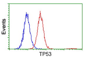
HEK293T cells transfected with either TP53 (Myc-DDK-tagged) overexpress plasmid (Red) or empty vector control plasmid (Blue) were immunostained by anti-TP53 antibody (M00001-5) |
|
|
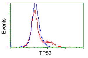
Flow cytometric Analysis of Hela cells |
|
|
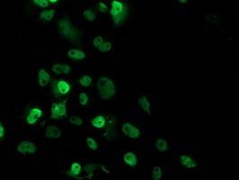
Anti-TP53 mouse monoclonal antibody (M00001-5) immunofluorescent staining of COS7 cells transiently transfected by pCMV6-ENTRY TP53. |
|
|
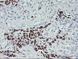
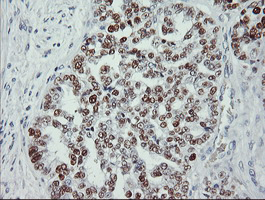
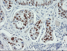
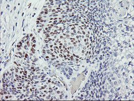
Immunohistochemical staining of paraffin-embedded Carcinoma of Human lung tissue using anti-TP53 mouse monoclonal antibody. (Heat-induced epitope retrieval by 10mM citric buffer |
|
|
Immunohistochemical staining of paraffin-embedded Adenocarcinoma of Human ovary tissue using anti-TP53 mouse monoclonal antibody. (Heat-induced epitope retrieval by 10mM citric buffer |
|
|
Immunohistochemical staining of paraffin-embedded Carcinoma of Human pancreas tissue using anti-TP53 mouse monoclonal antibody. (Heat-induced epitope retrieval by 10mM citric buffer |
|
|
Immunohistochemical staining of paraffin-embedded Carcinoma of Human bladder tissue using anti-TP53 mouse monoclonal antibody. (Heat-induced epitope retrieval by 10mM citric buffer |
|
|
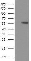
HEK293T cells were transfected with the pCMV6-ENTRY control (Left lane) or pCMV6-ENTRY TP53 (Right lane) cDNA for 48 hrs and lysed. Equivalent amounts of cell lysates (5 ug per lane) were separated by SDS-PAGE and immunoblotted with anti-TP53. |
|
|
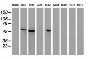
Western blot analysis of extracts (35ug) from 9 different cell lines by using anti-TP53 monoclonal antibody. |
Product Guarantee and Expert Support
