Rabbit IgG in stabilizing components, phosphate buffered saline, pH 7.4, 150mM NaCl, 0.02% sodium azide and 50% glycerol. *This antibody is supplied in a stabilized formulation. Compatibility with conjugation reactions depends on the chemistry of the con
Short-term DMOG treatment reduces MSC senescence by activating HIF-1alpha and decreasing apoptosis. ( A , B ) Representative images of SA-beta-gal staining showing the proportion of senescent cells in H2O2-treated MSCs before and after DMOG treatment. Scale bar =500µm. ( C ) Western blot analysis of p53 and p21 protein expression during oxidative stress-induced senescence. ( D , E ) SA-beta-gal staining in replicative senescence (P15) MSCs, demonstrating the effect of DMOG in reducing senescence markers. Scale bar =500µm. ( F ) Western blot analysis of p53 and p21 protein levels in P5 (young) and P15 (senescent) MSCs. ( G ) Expression levels of senescence-associated genes (IL6, CXCL1, and MMP3) in both senescence models as assessed by qRT-PCR. ( H ) Western blotting and qPCR analysis showing increased HIF-1alpha protein and mRNA levels in both senescence models after DMOG treatment. ( I ) Calcein/PI live-dead staining revealed an increase in the proportion of live cells following DMOG treatment in both senescence models. ( J , K ) Flow cytometric analysis of apoptosis. Error bars represent the meanSD of three independent experiments. Statistical significance was set as p <0.05. Full-length blots are presented in Supplementary Materials - WB Raw Data Full size imageIndex in PubMed under a CC BY license. PMID: 40457488
Activation of HIF-1alpha/BNIP3 signaling pathway in artery of KD murine model. A Representative immumohistochemical images for HIF-1alpha expression in aorta ( n =4 per group). Enlarged images of area of interesting (AOI) were indicated with red block diagrams. Scale bar: 50µm. B Representative immumohistochemical images for HIF-1alpha expression in coronary bifurcation arteritis ( n =4 per group). Scale bar: 50µm. C Representative immumohistochemical images for BNIP3 expression in aorta ( n =4 per group). Enlarged images of area of interesting (AOI) were indicated with red block diagrams. Scale bar: 50µm. D Representative immumohistochemical images for BNIP3 expression in coronary bifurcation arteritis ( n =4 per group). Scale bar: 50µm. Index in PubMed under a CC BY license. PMID: 37980402
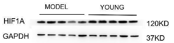
Western blot analysis of HIF-1 alpha using anti-HIF-1 alpha antibody (M00013-3). Electrophoresis was performed on a 5-20% SDS-PAGE gel at 80V (Stacking gel) / 120V (Resolving gel) for 2 hours. The sample well of each lane was loaded with 30 ug of sample under reducing conditions. Lane 1-5: model group-human uterine tissue lysates, Lane 6-10: young group-human uterine tissue- lysates. After electrophoresis, proteins were transferred to a nitrocellulose membrane at 150 mA for 50-90 minutes. Blocked the membrane with 5% non-fat milk/TBS for 1.5 hour at RT. The membrane was incubated with rabbit anti-HIF-1 alpha antigen affinity purified monoclonal antibody (A04887-1) at 1:1000 overnight at 4C, then washed with TBS-0.1%Tween 3 times with 5 minutes each and probed with a goat anti-rabbit IgG-HRP secondary antibody at for 1 hour at RT. The signal is developed using an ECL Plus Western Blotting Substrate (Catalog AR1196-200) with Tanon 5200 system. A specific band was detected for HIF-1 alpha at approximately 120 kDa. The expected band size for HIF-1 alpha is at 120 kDa.
Immunofluorescent analysis of Hela cells, using HIF-1 alpha Antibody.
Immunohistochemical analysis of paraffin-embedded human kindey, using HIF-1 alpha Antibody.
Western blot analysis of HIF-1 alpha expression in Ramos cell lysate.
All lanes use the Antibody at 1:1000 dilution for 1 hour at room temperature.
All lanes use the Antibody at 1:1000 dilution for 1 hour at room temperature.
All lanes use the Antibody at 1:1000 dilution for 1 hour at room temperature.
* VAT and and shipping costs not included. Errors and price changes excepted