Full length human recombinant protein of human TGFB1 (NP_000651) produced in HEK293T cell.
Boster Bio TGFB1 mouse monoclonal antibody, clone OTI3B6 (formerly 3B6). Catalog M00019-3. Tested in FC, IF, WB. This antibody reacts with Human, Mouse, Rat.
PBS (pH 7.3) containing 1% stabilizing protein, 50% glycerol and 0.02% sodium azide.This antibody is supplied in a stabilized formulation. Compatibility with conjugation reactions depends on the chemistry of the conjugation method used. For conjugation me
Application Dilute:
WB: 1:2000IF: 1:100
Lactucin exerts antifibrotic effects by impacting the expression of fibrosis-related factors. ( A - C ) The immunofluorescence assay was used to assess the potential effect on translocation of Stat3, Image Scale 50µm (n=3, SD, *P<0.05, **P<0.01), ( D - I ) The levels of TLR4, Smad3, Smad7, TGF-beta1 and alpha-SMA were determined by Western blot (n=3, SD, *P<0.05, **P<0.01). Index in PubMed under a CC BY license. PMID: 39164375
Lactucin exert antifibrotic effects in mice by affecting the expression of fibrosis-related factors. The levels of TLR4, Smad3, Smad7, TGF-beta1, alpha-SMA and Stat3 were determined by Western blot (n=3). The data are presented as the meansSD in each group. * P <0.05, ** P <0.01 with the Model Group, P <0.05, P <0.01 with the Normal Group. Normal healthy mice, Model mice with liver fibrosis, L-Lactucin Lactucin low-dose treatment group, M-Lactucin Lactucin middle-dose treatment group, H-Lactucin Lactucin high-dose treatment group. Index in PubMed under a CC BY license. PMID: 39164375
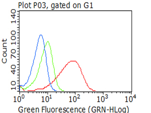
Flow cytometric Analysis of permeabilized HCT116 cells
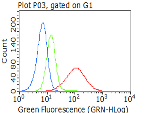
Flow cytometric Analysis of permeabilized SF295 cells
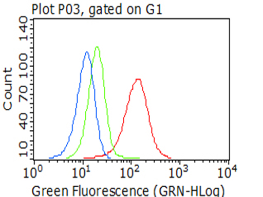
Flow cytometric Analysis of permeabilized Hela cells
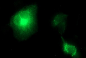
Anti-TGFB1 mouse monoclonal antibody (M00019-3) immunofluorescent staining of COS7 cells transiently transfected by pCMV6-ENTRY TGFB1 (1:100).
Immunofluorescent staining of HeLa cells using anti-TGFB1 mouse monoclonal antibody (M00019-3) (1:100).
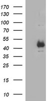
HEK293T cells were transfected with the pCMV6-ENTRY control (Left lane) or pCMV6-ENTRY TGFB1 (Right lane) cDNA for 48 hrs and lysed. Equivalent amounts of cell lysates (5 ug per lane) were separated by SDS-PAGE and immunoblotted with anti-TGFB1 (1:2000).
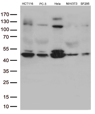
Western blot analysis of extracts (35ug) from 5 different cell lines by using anti-TGFB1 monoclonal antibody (1:500).
* VAT and and shipping costs not included. Errors and price changes excepted