BRD4, bromodomain containing 4, CAP, HUNK1, HUNKI, MCAP, Protein HUNK1
Boster Bio Anti-Brd4 Rabbit Monoclonal Antibody catalog M00123. Tested in WB, IHC, ICC/IF, IP applications. This antibody reacts with Human, Mouse, Rat.
Rabbit IgG in stabilizing components, phosphate buffered saline, pH 7.4, 150mM NaCl, 0.02% sodium azide and 50% glycerol. *This antibody is supplied in a stabilized formulation. Compatibility with conjugation reactions depends on the chemistry of the con
Purity:
Affinity-chromatography
Form:
Liquid
Target:
Bromodomain-containing protein 4
Application Dilute:
WB 1:500-2000IHC 1:50-200ICC/IF 1:50-200IP 1:20
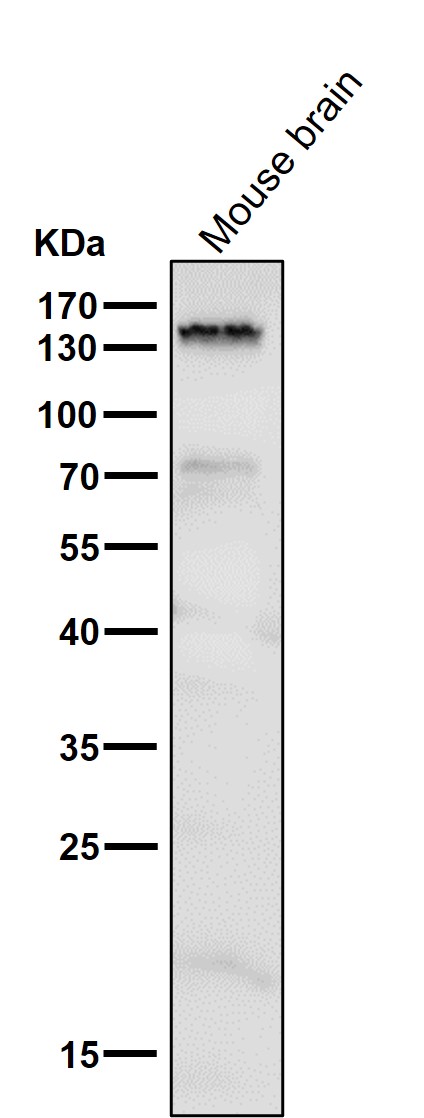
Western blot analysis of Brd4 using anti-Brd4 antibody (M00123). Electrophoresis was performed on a 8% SDS-PAGE gel at 70V (Stacking gel) / 90V (Resolving gel) for 2-3 hours. The sample well of each lane was loaded with 30 ug of sample under reducing conditions. Lane 1: mouse brain tissue lysates. After electrophoresis, proteins were transferred to a nitrocellulose membrane at 150 mA for 50-90 minutes. Blocked the membrane with 5% non-fat milk/TBS for 1.5 hour at RT. The membrane was incubated with rabbit anti-Brd4 antigen affinity purified monoclonal antibody (M05837-1) at 1:500 overnight at 4C, then washed with TBS-0.1%Tween 3 times with 5 minutes each and probed with a goat anti-rabbit IgG-HRP secondary antibody at a dilution of 1:500 for 1.5 hour at RT. The signal is developed using an Enhanced Chemiluminescent detection (ECL) kit (Catalog AR1196-200) with Tanon 5200 system. A specific band was detected for Brd4 at approximately 240 kDa. The expected band size for Brd4 is at 152 kDa.
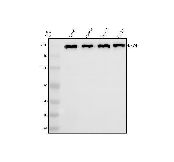
Western blot analysis of Brd4 using anti-Brd4 antibody (M00123). Electrophoresis was performed on a 8% SDS-PAGE gel at 70V (Stacking gel) / 90V (Resolving gel) for 2-3 hours. The sample well of each lane was loaded with 30 ug of sample under reducing conditions. Lane 1: human Jurkat whole cell lysates,Lane 2: human HepG2 whole cell lysates,Lane 3: human MCF-7 whole cell lysates,Lane 4: human PC-12 whole cell lysates.After electrophoresis, proteins were transferred to a nitrocellulose membrane at 150 mA for 50-90 minutes. Blocked the membrane with 5% non-fat milk/TBS for 1.5 hour at RT. The membrane was incubated with rabbit anti-Brd4 antigen affinity purified monoclonal antibody (M05837-1) at 1:500 overnight at 4C, then washed with TBS-0.1%Tween 3 times with 5 minutes each and probed with a goat anti-rabbit IgG-HRP secondary antibody at a dilution of 1:500 for 1.5 hour at RT. The signal is developed using an Enhanced Chemiluminescent detection (ECL) kit (Catalog AR1196-200) with Tanon 5200 system. A specific band was detected for Brd4 at approximately 240 kDa. The expected band size for Brd4 is at 152 kDa.
Immunofluorescent analysis of Hela cells, using Brd4 Antibody .
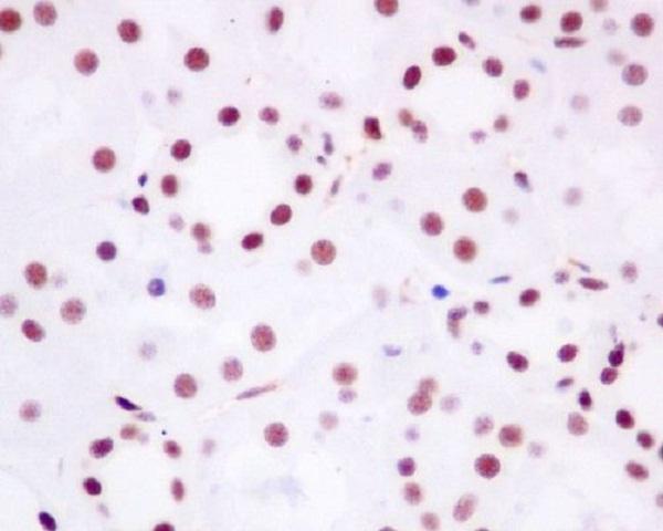
Immunohistochemical analysis of paraffin-embedded human kidney, using Brd4 Antibody.
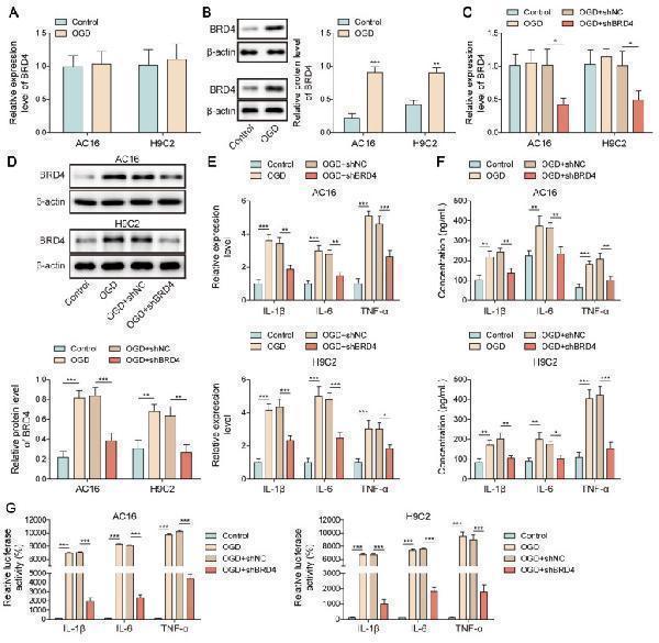
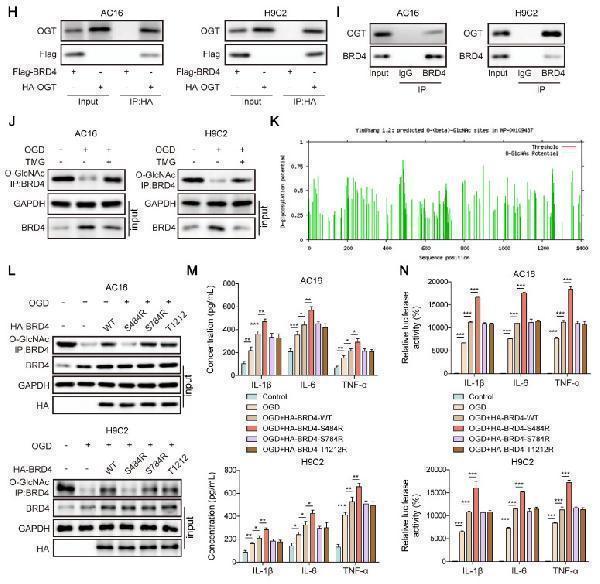
O-GlcNAcylation of BRD4 inhibited NF-kappaB p65-mediated transcription of pro-inflammatory cytokines. (A)&(B) The expression of BRD4 in OGD-exposed cardiomyocytes was detected by RT-qPCR and Western blotting. H9C2 and AC-16 cells were transfected with shBRD4, and then subjected to OGD. (C)&(D) RT-qPCR and Western blotting analysis of BRD4 mRNA and protein levels. (E)&(F) The mRNA levels and concentrations of TNF-alpha, IL-1beta, and IL-6 were determined by RT-qPCR and ELISA. (G) The binding of NF-kappaB p65 to TNF-alpha, IL-1beta, and IL-6 promoters was confirmed by dual-luciferase reporter assay. (H)&(I) Co-IP assay verified the exogenous and endogenous interplay between OGT and BRD4 proteins. (J) O-GlcNAcylation of BRD4 protein in OGD-stimulated cardiomyocytes was evaluated. (K) YinOYang database predicated the potential O-GlcNAc sites on BRD4. OGD-challenged H9C2 and AC-16 cells were transfected with BRD4 WT plasmid or BRD4 plasmids with mutant O-GlcNAc sites (BRD4-S484R, BRD4-S784R, and BRD4-T1212R). (L) O-GlcNAcylation of BRD4 protein in H9C2 and AC-16 cells was detected. (M) Concentrations of TNF-alpha, IL-1beta, and IL-6 were detected by ELISA. (N) The interaction between NF-kappaB p65 and TNF-alpha, IL-1beta, and IL-6 promoters was validated by dual-luciferase reporter assay. n=3 for A-N. Students t test (for A, B) and one-way ANOVA (for C-G, M, N) were performed to analyze data. * p < 0.05, ** p < 0.01, *** p < 0.001.Index in PubMed under a CC BY license. PMID: 40585977
O-GlcNAcylation of BRD4 inhibited NF-kappaB p65-mediated transcription of pro-inflammatory cytokines. (A)&(B) The expression of BRD4 in
* VAT and and shipping costs not included. Errors and price changes excepted