CDK2, Cell division protein kinase 2, cyclin dependent kinase 2, p33 protein kinase, p33(CDK2)
Boster Bio Anti-CDK2 Rabbit Monoclonal Antibody catalog M00166. Tested in WB, IHC, ICC/IF, IP applications. This antibody reacts with Human, Mouse, Rat.
Rabbit IgG in stabilizing components, phosphate buffered saline, pH 7.4, 150mM NaCl, 0.02% sodium azide and 50% glycerol. *This antibody is supplied in a stabilized formulation. Compatibility with conjugation reactions depends on the chemistry of the con
Purity:
Affinity-chromatography
Form:
Liquid
Target:
Cyclin-dependent kinase 2
Application Dilute:
WB 1:500-2000IHC 1:50-200ICC/IF 1:50-200IP 1:30
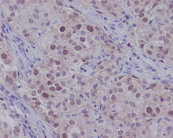
Immunohistochemical analysis of paraffin-embedded human beast carcinoma, using CDK2 Antibody.
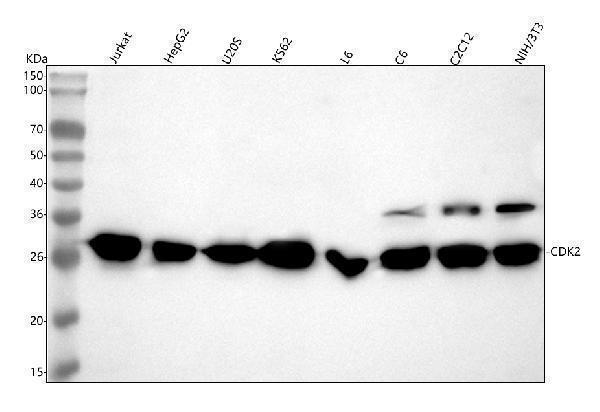
Western blot analysis of CDK2 using anti-CDK2 antibody (M00166). Electrophoresis was performed on a 5-20% SDS-PAGE gel at 70V (Stacking gel) / 90V (Resolving gel) for 2-3 hours. The sample well of each lane was loaded with 30 ug of sample under reducing conditions. Lane 1: human Jurkat whole cell lysates,Lane 2: human HepG2 whole cell lysates,Lane 3: human U2OS whole cell lysates,Lane 4: human K562 whole cell lysates,Lane 5: rat L6 whole cell lysates,Lane 6: rat C6 whole cell lysates,Lane 7: mouse C2C12 whole cell lysates,Lane 8: mouse NIH/3T3 whole cell lysates.After electrophoresis, proteins were transferred to a nitrocellulose membrane at 150 mA for 50-90 minutes. Blocked the membrane with 5% non-fat milk/TBS for 1.5 hour at RT. The membrane was incubated with rabbit anti-CDK2 antigen affinity purified monoclonal antibody (Catalog M00166) at 1:500 overnight at 4C, then washed with TBS-0.1%Tween 3 times with 5 minutes each and probed with a goat anti-rabbit IgG-HRP secondary antibody at a dilution of 1:1000 for 1.5 hour at RT. The signal is developed using an Enhanced Chemiluminescent detection (ECL) kit (Catalog EK1002) with Tanon 5200 system. A specific band was detected for CDK2 at approximately 30 kDa. The expected band size for CDK2 is at 34 kDa.
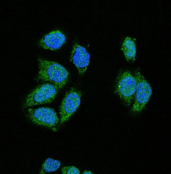
Immunofluorescent analysis using the Antibody at 1:150 dilution.
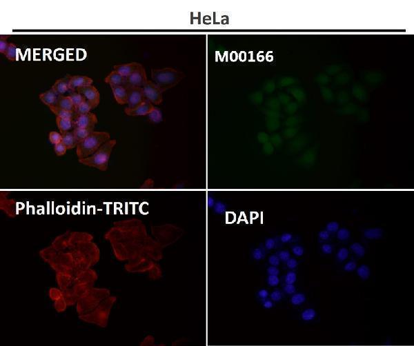
Immunofluorescent analysis of Hela cells, using CDK2 Antibody .
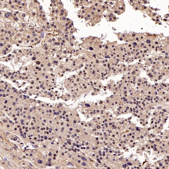
Immunohistochemical analysis of paraffin-embedded Human liver cancer, using the Antibody at 1:100 dilution.
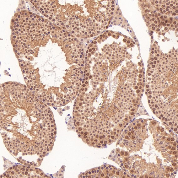
Immunohistochemical analysis of paraffin-embedded Mouse testis, using the Antibody at 1:100 dilution.
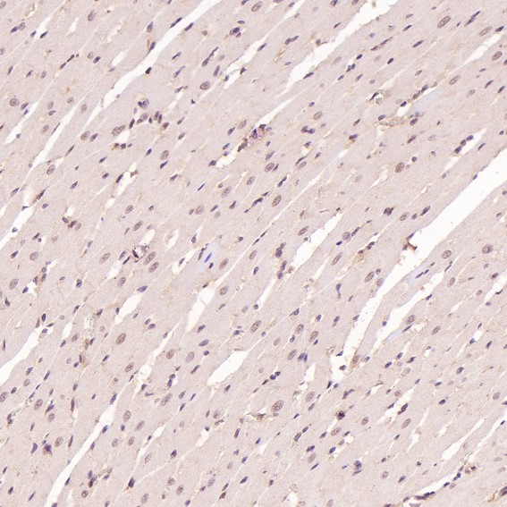
Immunohistochemical analysis of paraffin-embedded Rat heart, using the Antibody at 1:200 dilution.
Immunohistochemical analysis of paraffin-embedded Rat pancreas, using the Antibody at 1:200 dilution.
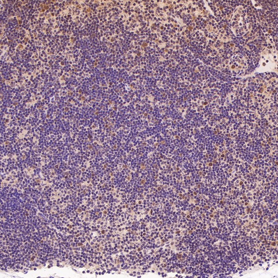
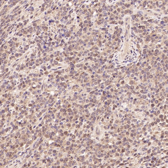
Immunohistochemical analysis of paraffin-embedded Human non-Hodgkins lymphoma, using the Antibody at 1:100 dilution.
* VAT and and shipping costs not included. Errors and price changes excepted