
CCR7 Rabbit Polyclonal Antibody, Unconjugated
Catalog Number:
BYT-ORB10276
- Images (9)


| Article Name: | CCR7 Rabbit Polyclonal Antibody, Unconjugated |
| Biozol Catalog Number: | BYT-ORB10276 |
| Supplier Catalog Number: | orb10276 |
| Alternative Catalog Number: | BYT-ORB10276-100,BYT-ORB10276-200,BYT-ORB10276-50 |
| Manufacturer: | Biorbyt |
| Host: | Rabbit |
| Category: | Antikörper |
| Application: | FC, ICC, IF, IHC-Fr, IHC-P, WB |
| Species Reactivity: | Human, Mouse, Rat |
| Immunogen: | KLH conjugated synthetic peptide derived from human CCR7 (25-59/379aa) |
| Conjugation: | Unconjugated |
| Alternative Names: | CCR7_HUMAN, BLR 2, BLR2, C C chemokine receptor type 7, C C CKR 7, CC chemokine receptor 7, CC chemokine receptor type 7, CC CKR 7, CCCKR7, CCR 7, CD 197, CD197, CD197 antigen, CDW197, Chemokine C C motif receptor 7, Chemokine C C receptor 7, Chemokine receptor 7-like protein, EBI 1, EBI1, Ebi1h, EBV Induced G Protein Coupled Receptor 1, Epstein Barr virus induced G protein coupled receptor, Epstein Barr virus induced gene 1, EVI 1, EVI1, Lymphocyte Specific G Protein Coupled Peptide Receptor, MGC108519, MIP 3 beta receptor, MIP3 beta Receptor. |
| CCR7 Rabbit Polyclonal Antibody |
| Application Dilute: | WB=1:500-2000, IHC-P=1:100-500, IHC-F=1:100-500, ICC/IF=1:100-500, IF=1:100-500, Flow-Cyt=1µg/Test |
|
|
Blank control: THP-1. Primary Antibody (green line): Rabbit Anti-CCR7 antibody (orb10276), Dilution: 1 µg/10 6 cells, Isotype Control Antibody (orange line): Rabbit IgG. Secondary Antibody: Goat anti-rabbit IgG-FITC, Dilution: 0.5 µg/test. Protocol, The cells were fixed with 4% PFA (10 min at room temperature) and then permeabilized with 0.1% PBST for 20 min at room temperature. The cells were then incubated in 5% BSA to block non-specific protein-protein interactions for 30 min at room temperature. Cells stained with Primary Antibody for 30 min at room temperature. The secondary antibody used for 40 min at room temperature. Acquisition of 20000 events was performed. |
|
|
Hela cell, 4% Paraformaldehyde-fixed, Triton X-100 at room temperature for 20 min, Blocking buffer (normal goat serum) at 37C for 20 min, Antibody incubation with (CCR7) polyclonal Antibody, Unconjugated (orb10276) 1:100, 90 minutes at 37C, followed by a conjugated Goat Anti-Rabbit IgG antibody at 37C for 90 minutes, DAPI (blue) was used to stain the cell nuclei. |
|
|
Paraformaldehyde-fixed, paraffin embedded (RAT lymphoid), Antigen retrieval by boiling in sodium citrate buffer (pH6.0) for 15 min, Block endogenous peroxidase by 3% hydrogen peroxide for 20 minutes, Blocking buffer (normal goat serum) at 37C for 30 min, Antibody incubation with (CCR7) Polyclonal Antibody, Unconjugated (orb10276) at 1:200 overnight at 4C, followed by operating according to SP Kit (Rabbit) instructionsand DAB staining. |
|
|
Sample: Lane 1: Mouse Bone tissue lysates, Lane 2: Mouse Raw264.7 cell lysates, Lane 3: Human Jurkat cell lysates, Lane 4: Human MCF-7 cell lysates, Lane 5: Human THP-1 cell lysates, Primary: Anti-CCR7 (orb10276) at 1/1000 dilution, Secondary: IRDye800CW Goat Anti-Rabbit IgG at 1/20000 dilution, Predicted band size: 42 kDa, Observed band size: 42 kDa. |
|
|
Tissue/cell: human gastric tissue, 4% Paraformaldehyde-fixed and paraffin-embedded, Antigen retrieval: citrate buffer (0.01M, pH 6.0), Boiling bathing for 15 min, Blocking buffer (normal goat serum) at 37°C for 20 min, Incubation: Anti-CCR7 Polyclonal Antibody, Unconjuga |
|
|
|
|
|
Hela cell, 4% Paraformaldehyde-fixed, Triton X-100 at room temperature for 20 min, Blocking buffer (normal goat serum) at 37C for 20 min, Antibody incubation with (CCR7) polyclonal Antibody, Unconjugated (orb10276) 1:100, 90 minutes at 37C, followed by a conjugated Goat Anti-Rabbit IgG antibody at 37C for 90 minutes, DAPI (blue) was used to stain the cell nuclei. |
|
|
Blank control (Black line): Mouse spleen (Black). Primary Antibody (green line): Rabbit Anti-CD4 antibody, Dilution: 3 µg/10 6 cells, Isotype Control Antibody (orange line): Rabbit IgG-PE. Secondary Antibody (white blue line): Goat anti-rabbit IgG-PE, Dilution: 1 µg/test. Protocol, The cells were fixed with 4% PFA (10 min at room temperature) and then permeabilized with 90% ice-cold methanol for 20 min at room temperature. The cells were then incubated in 5% BSA to block non-specific protein-protein interactions for 30 min at room temperature. Cells stained with Primary Antibody for 30 min at room temperature. The secondary antibody used for 40 min at room temperature. Acquisition of 20000 events was performed. |
|
|
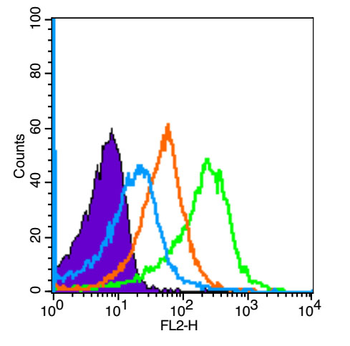
Blank control: Raji (blue). Primary Antibody: Rabbit Anti-CCR7 antibody (orb10276), Dilution: 1 µg in 100 µL 1X PBS containing 0.5% BSA, Isotype Control Antibody: Rabbit IgG (orange), used under the same conditions, Secondary Antibody: Goat anti-rabbit IgG-PE (white blue), Dilution: 1:200 in 1 X PBS containing 0.5% BSA. Protocol, The cells were fixed with 2% paraformaldehyde (10 min). Primary antibody (orb10276, 1 µg/1x10 6 cells) were incubated for 30 min on the ice, followed by 1 X PBS containing 0.5% BSA + 10% goat serum (15 min) to block non-specific protein-protein interactions. Then the Goat Anti-rabbit IgG/PE antibody was added into the blocking buffer mentioned above to react with the primary antibody at 1/200 dilution for 30 min on ice. Acquisition of 20000 events was performed. |
Product Guarantee and Expert Support
