Supplied at 0.5 mg/ml in Tris saline, 0.02% sodium azide, pH 7.3 with 0.5% bovine serum albumin. Aliquot and store at -20C. Minimize freezing and thawing.
Sequence:
SEIQYKILTQKED
Target:
IFNGR1 (aa181-193)
Application Dilute:
ELISA: 1:128000, WB: 0.3-1 µg/ml
Application Notes:
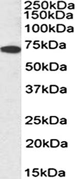
Application Notes: WB: Approx 70kDa band observed in lysates of cell line HepG2 (calculated MW of 54.4kDa according to NP_000407.1). The observed molecular weight corresponds to the glycosylated form. Recommended concentration: 0.3-1µg/ml
Western blot analysis of HepG2 lysate (35ug protein in RIPA buffer) using IFNGR1 antibody
1 µg/ml staining of K562 (A) Caco-2 (B) and HepG2 (C) cell lysate (35 µg protein in RIPA buffer). Detected by chemiluminescence.
2 µg/ml staining of Human Spleen lysate (35 µg protein in RIPA buffer). Detected by chemiluminescence.
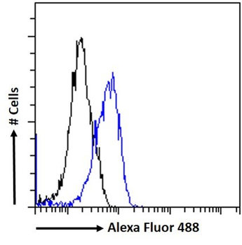
Flow cytometric analysis of paraformaldehyde fixed K562 cells (blue line), permeabilized with 0.5% Triton. Primary incubation 1hr (10 ug/ml) followed by Alexa Fluor 488 secondary antibody (1 ug/ml). IgG control: Unimmunized goat IgG (black line) followed by Alexa Fluor 488 secondary antibody.
Immunofluorescence analysis of paraformaldehyde fixed Caco-2 cells, permeabilized with 0.15% Triton. Primary incubation 1hr (10 ug/ml) followed by Alexa Fluor 488 secondary antibody (2 ug/ml), showing membrane staining. The nuclear stain is DAPI (blue). Negative control: Unimmunized goat IgG (10 ug/ml) followed by Alexa Fluor 488 secondary antibody (2 ug/ml).
Immunofluorescence analysis of paraformaldehyde fixed THP-1 cells immobilized on ShifixTM coverslip, permeabilized with 0.15% Triton. Primary incubation 1hr (10 ug/ml) followed by Alexa Fluor 488 secondary antibody (2 ug/ml), showing membrane and cytoplasmic staining. The nuclear stain is DAPI (blue). Negative control: Unimmunized goat IgG (10 ug/ml) followed by Alexa Fluor 488 secondary antibody (2 ug/ml).
4 µg/ml staining of paraffin embedded Human Lung. Heat induced antigen retrieval with citrate buffer pH6, HRP-staining.
Negative Control showing staining of paraffin embedded Human Lung, with no primary antibody.
* VAT and and shipping costs not included. Errors and price changes excepted