
HSP27 Rabbit Polyclonal Antibody, Unconjugated
Catalog Number:
BYT-ORB10845
- Images (8)


| Article Name: | HSP27 Rabbit Polyclonal Antibody, Unconjugated |
| Biozol Catalog Number: | BYT-ORB10845 |
| Supplier Catalog Number: | orb10845 |
| Alternative Catalog Number: | BYT-ORB10845-50,BYT-ORB10845-100,BYT-ORB10845-200 |
| Manufacturer: | Biorbyt |
| Host: | Rabbit |
| Category: | Antikörper |
| Application: | FC, IF, IHC-Fr, IHC-P, WB |
| Species Reactivity: | Human, Rat |
| Immunogen: | KLH conjugated synthetic peptide derived from human HSP27 (101-205/205aa) |
| Conjugation: | Unconjugated |
| Alternative Names: | CMT2F, HEL-S-102, HMN2B, HMND3, HS.76067, HSP27, HSP28, Hsp25, SRP27, 27kDa, HSPB1_CANLF, HSPB1, Heat shock 27 kDa protein (HSP 27), HSPB1_HUMAN, 28 kDa heat shock protein, Estrogen-regulated 24 kDa protein, Heat shock protein family B member 1, Stress-responsive protein 27 (SRP27), HSPB1_MOUSE, Growth-related 25 kDa protein, Heat shock 25 kDa protein (HSP 25), p25, HSPB1_RAT, |
| HSP27 Rabbit Polyclonal Antibody |
| Clonality: | Polyclonal |
| Concentration: | 1mg/ml |
| Molecular Weight: | 27 kDa |
| UniProt: | P04792 |
| Buffer: | 0.01M TBS (pH7.4) with 1% rAlbumin, 0.02% Proclin300 and 50% Glycerol. |
| Form: | Liquid |
| Target: | HSPB1 |
| Application Dilute: | WB=1:500-2000, IHC-P=1:100-500, IHC-F=1:100-500, IF=1:100-500, Flow-Cyt=2µg/Test |
|
|
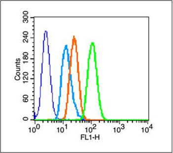
Blank control (blue line): A431 cells (blue). Primary Antibody (green line): Rabbit Anti-HSP27 antibody (orb10845), Dilution: 2 µg/10 6 cells, Isotype Control Antibody (orange line): Rabbit IgG. Secondary Antibody (white blue line): Goat anti-rabbit IgG-FITC, Dilution: 1 µg/Test. Protocol, The cells were fixed with 70% methanol (Overnight at 4C) and then permeabilized with 90% ice-cold methanol for 20 min at -20C. Cells stained with Primary Antibody for 30 min at room temperature. The cells were then incubated in 1 X PBS/2% BSA/10% goat serum to block non-specific protein-protein interactions followed by the antibody for 15 min at room temperature. The secondary antibody used for 40 min at room temperature. Acquisition of 20000 events was performed. |
|
|
Paraformaldehyde-fixed, paraffin embedded (Human breast cancer), Antigen retrieval by boiling in sodium citrate buffer (pH6.0) for 15 min, Block endogenous peroxidase by 3% hydrogen peroxide for 20 minutes, Blocking buffer (normal goat serum) at 37C for 30 min, Antibody incubation with (HSP27) Polyclonal Antibody, Unconjugated (orb10845) at 1:400 overnight at 4C, followed by operating according to SP Kit (Rabbit) instructions and DAB staining. |
|
|
Paraformaldehyde-fixed, paraffin embedded (rat skeletal muscle), Antigen retrieval by boiling in sodium citrate buffer (pH6.0) for 15 min, Block endogenous peroxidase by 3% hydrogen peroxide for 20 minutes, Blocking buffer (normal goat serum) at 37C for 30 min, Antibody incubation with (HSP27) Polyclonal Antibody, Unconjugated (orb10845) at 1:200 overnight at 4C, followed by operating according to SP Kit (Rabbit) instructionsand DAB staining. |
|
|
Paraformaldehyde-fixed, paraffin embedded (rat stomach tissue), Antigen retrieval by boiling in sodium citrate buffer (pH6.0) for 15 min, Block endogenous peroxidase by 3% hydrogen peroxide for 20 minutes, Blocking buffer (normal goat serum) at 37C for 30 min, Antibody incubation with (HSP27) Polyclonal Antibody, Unconjugated (orb10845) at 1:400 overnight at 4C, followed by operating according to SP Kit (Rabbit) instructionsand DAB staining. |
|
|
Paraformaldehyde-fixed, paraffin embedded (Rat uterus), Antigen retrieval by boiling in sodium citrate buffer (pH6.0) for 15 min, Block endogenous peroxidase by 3% hydrogen peroxide for 20 minutes, Blocking buffer (normal goat serum) at 37C for 30 min, Antibody incubation with (HSP27) Polyclonal Antibody, Unconjugated (orb10845) at 1:400 overnight at 4C, followed by operating according to SP Kit (Rabbit) instructionsand DAB staining. |
|
|
Sample: Brain (Mouse) lysate at 30 ug, Liver (Mouse) lysate at 30 ug, Primary: Anti-HSP-27 (orb10845) at 1:200, Secondary: AP conjugated Goat Anti-Rabbit IgG at 1:3000 dilution, NBT/BCIP staining, Predicted band size: 27kD, Observed band size: 27kD. |
|
|
Sample: Lane 1: Hela (Human) Cell Lysate at 30 ug, Lane 2: MCF-7 (Human) Cell Lysate at 30 ug, Lane 3: A431 (Human) Cell Lysate at 30 ug, Lane 4: Huvec (Human) Cell Lysate at 30 ug, Lane 5: HepG2 (Human) Cell Lysate at 30 ug, Primary: Anti-HSP27 (orb10845) at 1/1000 dilution, Secondary: IRDye800CW Goat Anti-Rabbit IgG at 1/20000 dilution, Predicted band size: 27-30 kD, Observed band size: 27 kD. |
|
|
Tissue/Cell: rat brain tissue, 4% Paraformaldehyde-fixed and paraffin-embedded, Antigen retrieval: citrate buffer (0.01M, pH6.0), Boiling bathing for 15 min, Blocking buffer (normal goat serum) at 37C for 20 min, Incubation: Anti-HSP-27 Polyclonal Antibody, Unconjugated (orb10845) 1:200, overnight at 4C, The secondary antibody was Goat Anti-Rabbit IgG, Cy3 conjugated (orb868589) used at 1:200 dilution for 40 minutes at 37C. DAPI (5 ug/ml, blue) was used to stain the cell nuclei. |
Product Guarantee and Expert Support
