A synthetic peptide corresponding to a sequence in the middle region of human G3BP/G3BP1, which shares 64.7% amino acid (aa) sequence identity with mouse G3BP1.
Conjugation:
Unconjugated
Alternative Names:
G3BP, G3BP 1, G3BP1, HDH VIII
Anti-G3BP/G3BP1 Antibody. Tested in Flow Cytometry, IF, IHC, ICC, WB applications. This antibody reacts with Human.
Clonality:
Polyclonal
Concentration:
Adding 0.2 ml of distilled water will yield a concentration of 500 µg/ml.
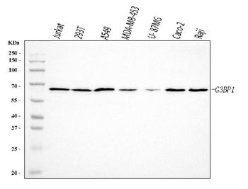
Western blot, 0.1-0.25 µg/ml, Human Immunohistochemistry(Paraffin-embedded Section), 2-5 µg/ml, Human Immunocytochemistry/Immunofluorescence, 5 µg/ml, Human Flow Cytometry (Fixed), 1-3 µg/1x10 6 cells, Human
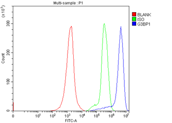
Flow Cytometry analysis of Jurkat cells using anti-G3BP/G3BP1 antibody. Overlay histogram showing Jurkat cells (Blue line). To facilitate intracellular staining, cells were fixed with 4% paraformaldehyde and permeabilized with permeabilization buffer. The cells were blocked with 10% normal goat serum. And then incubated with rabbit anti-G3BP/G3BP1 Antibody (1 µg/1x10 6 cells) for 30 min at 20C. DyLight488 conjugated goat anti-rabbit IgG (5-10 µg/1x10 6 cells) was used as secondary antibody for 30 minutes at 20C. Isotype control antibody (Green line) was rabbit IgG (1 µg/1x10 6) used under the same conditions. Unlabelled sample without incubation with primary antibody and secondary antibody (Red line) was used as a blank control.
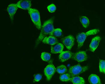
IF analysis of G3BP/G3BP1 using anti-G3BP/G3BP1 antibody. G3BP/G3BP1 was detected in an immunocytochemical section of Caco-2 cells. Enzyme antigen retrieval was performed using IHC enzyme antigen retrieval reagent for 15 mins. The cells were blocked with 10% goat serum. And then incubated with 5 µg/mL rabbit anti-G3BP/G3BP1 Antibody overnight at 4C. DyLight488 Conjugated Goat Anti-Rabbit IgG was used as secondary antibody at 1:100 dilution and incubated for 30 minutes at 37C. The section was counterstained with DAPI. Visualize using a fluorescence microscope and filter sets appropriate for the label used.
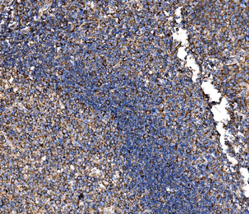
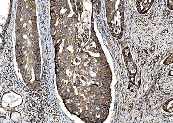
IHC analysis of G3BP/G3BP1 using anti-G3BP/G3BP1 antibody. G3BP/G3BP1 was detected in a paraffin-embedded section of human adenocarcinoma of the right colon tissue. Heat mediated antigen retrieval was performed in EDTA buffer (pH8.0, epitope retrieval solution). The tissue section was blocked with 10% goat serum. The tissue section was then incubated with 2 µg/ml rabbit anti-G3BP/G3BP1 Antibody overnight at 4C. Peroxidase Conjugated Goat Anti-rabbit IgG was used as secondary antibody and incubated for 30 minutes at 37C. The tissue section was developed using HRP Conjugated Rabbit IgG Super Vision Assay Kit with DAB as the chromogen.
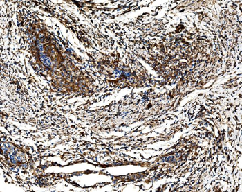
IHC analysis of G3BP/G3BP1 using anti-G3BP/G3BP1 antibody. G3BP/G3BP1 was detected in a paraffin-embedded section of human cervical cancer tissue. Heat mediated antigen retrieval was performed in EDTA buffer (pH8.0, epitope retrieval solution). The tissue section was blocked with 10% goat serum. The tissue section was then incubated with 2 µg/ml rabbit anti-G3BP/G3BP1 Antibody overnight at 4C. Peroxidase Conjugated Goat Anti-rabbit IgG was used as secondary antibody and incubated for 30 minutes at 37C. The tissue section was developed using HRP Conjugated Rabbit IgG Super Vision Assay Kit with DAB as the chromogen.
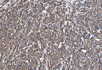
IHC analysis of G3BP/G3BP1 using anti-G3BP/G3BP1 antibody. G3BP/G3BP1 was detected in a paraffin-embedded section of human liver cancer tissue. Heat mediated antigen retrieval was performed in EDTA buffer (pH8.0, epitope retrieval solution). The tissue section was blocked with 10% goat serum. The tissue section was then incubated with 2 µg/ml rabbit anti-G3BP/G3BP1 Antibody overnight at 4C. Peroxidase Conjugated Goat Anti-rabbit IgG was used as secondary antibody and incubated for 30 minutes at 37C. The tissue section was developed using HRP Conjugated Rabbit IgG Super Vision Assay Kit with DAB as the chromogen.
IHC analysis of G3BP/G3BP1 using anti-G3BP/G3BP1 antibody. G3BP/G3BP1 was detected in a paraffin-embedded section of human lung cancer tissue. Heat mediated antigen retrieval was performed in EDTA buffer (pH8.0, epitope retrieval solution). The tissue section was blocked with 10% goat serum. The tissue section was then incubated with 2 µg/ml rabbit anti-G3BP/G3BP1 Antibody overnight at 4C. Peroxidase Conjugated Goat Anti-rabbit IgG was used as secondary antibody and incubated for 30 minutes at 37C. The tissue section was developed using HRP Conjugated Rabbit IgG Super Vision Assay Kit with DAB as the chromogen.
IHC analysis of G3BP/G3BP1 using anti-G3BP/G3BP1 antibody. G3BP/G3BP1 was detected in a paraffin-embedded section of human lymphom
IHC analysis of G3BP/G3BP1 using anti-G3BP/G3BP1 antibody. G3BP/G3BP1 was detected in a p
* VAT and and shipping costs not included. Errors and price changes excepted