E.coli-derived human MSH2 recombinant protein (Position: Q337-N583). Human MSH2 shares 94% and 93% amino acid (aa) sequence identity with mouse and rat MSH2, respectively.
Each vial contains 4 mg Trehalose, 0.9 mg NaCl and 0.2 mg Na2HPO4.
Form:
Lyophilized
Target:
DNA mismatch repair protein Msh2
Application Dilute:
Western blot, 0.25-0.5 µg/ml, Human Immunohistochemistry(Paraffin-embedded Section), 2-5 µg/ml, Human Immunocytochemistry/Immunofluorescence, 5 µg/ml, Human
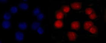
IF analysis of MSH2 using anti-MSH2 antibody. MSH2 was detected in an immunocytochemical section of Caco-2 cells. Enzyme antigen retrieval was performed using IHC enzyme antigen retrieval reagent for 15 mins. The cells were blocked with 10% goat serum. And then incubated with 5 µg/mL mouse anti-MSH2 Antibody overnight at 4C. DyLight594 Conjugated Goat Anti-Mouse IgG was used as secondary antibody at 1:100 dilution and incubated for 30 minutes at 37C. The section was counterstained with DAPI. Visualize using a fluorescence microscope and filter sets appropriate for the label used.
IHC analysis of MSH2 using anti-MSH2 antibody. MSH2 was detected in a paraffin-embedded section of human colorectal adenocarcinoma tissue. Heat mediated antigen retrieval was performed in EDTA buffer (pH8.0, epitope retrieval solution). The tissue section was blocked with 10% goat serum. The tissue section was then incubated with 2 µg/ml mouse anti-MSH2 Antibody overnight at 4C. Peroxidase Conjugated Goat Anti-mouse IgG was used as secondary antibody and incubated for 30 minutes at 37C. The tissue section was developed using HRP Conjugated Mouse IgG Super Vision Assay Kit with DAB as the chromogen.
IHC analysis of MSH2 using anti-MSH2 antibody. MSH2 was detected in a paraffin-embedded section of human invasive urothelial carcinoma tissue. Heat mediated antigen retrieval was performed in EDTA buffer (pH8.0, epitope retrieval solution). The tissue section was blocked with 10% goat serum. The tissue section was then incubated with 2 µg/ml mouse anti-MSH2 Antibody overnight at 4C. Peroxidase Conjugated Goat Anti-mouse IgG was used as secondary antibody and incubated for 30 minutes at 37C. The tissue section was developed using HRP Conjugated Mouse IgG Super Vision Assay Kit with DAB as the chromogen.
IHC analysis of MSH2 using anti-MSH2 antibody. MSH2 was detected in a paraffin-embedded section of human laryngeal squamous cell carcinomas tissue. Heat mediated antigen retrieval was performed in EDTA buffer (pH8.0, epitope retrieval solution). The tissue section was blocked with 10% goat serum. The tissue section was then incubated with 2 µg/ml mouse anti-MSH2 Antibody overnight at 4C. Peroxidase Conjugated Goat Anti-mouse IgG was used as secondary antibody and incubated for 30 minutes at 37C. The tissue section was developed using HRP Conjugated Mouse IgG Super Vision Assay Kit with DAB as the chromogen.
IHC analysis of MSH2 using anti-MSH2 antibody. MSH2 was detected in a paraffin-embedded section of human liver cancer tissue. Heat mediated antigen retrieval was performed in EDTA buffer (pH8.0, epitope retrieval solution). The tissue section was blocked with 10% goat serum. The tissue section was then incubated with 2 µg/ml mouse anti-MSH2 Antibody overnight at 4C. Peroxidase Conjugated Goat Anti-mouse IgG was used as secondary antibody and incubated for 30 minutes at 37C. The tissue section was developed using HRP Conjugated Mouse IgG Super Vision Assay Kit with DAB as the chromogen.
IHC analysis of MSH2 using anti-MSH2 antibody. MSH2 was detected in a paraffin-embedded section of human serous adenocarcinoma of ovary tissue. Heat mediated antigen retrieval was performed in EDTA buffer (pH8.0, epitope retrieval solution). The tissue section was blocked with 10% goat serum. The tissue section was then incubated with 2 µg/ml mouse anti-MSH2 Antibody overnight at 4C. Peroxidase Conjugated Goat Anti-mouse IgG was used as secondary antibody and incubated for 30 minutes at 37C. The tissue section was developed using HRP Conjugated Mouse IgG Super Vision Assay Kit with DAB as the chromogen.
IHC analysis of MSH2 using anti-MSH2 antibody. MSH2 was detected in a paraffin-embedded section of human tonsil tissue. Heat mediated antigen retrieval was performed in EDTA buffer (pH8.0, epitope retrieval solution). The tissue section was blocked with 10% goat serum. The tissue section was then incubated with 2 µg/ml mouse anti-MSH2 Antibody overnight at 4C.
* VAT and and shipping costs not included. Errors and price changes excepted