A synthetic peptide corresponding to a sequence at the C-terminus of human Ataxin 1, different from the related mouse and rat sequences by one amino acid.
Conjugation:
Unconjugated
Alternative Names:
ataxin 1, ATX1, D6S504E, SCA1, ATXN1
Anti-Ataxin 1 Antibody (monoclonal, 2B13G8). Tested in Flow Cytometry, IHC, WB applications. This antibody reacts with Human, Mouse, Rat.
Clonality:
Monoclonal
Concentration:
Adding 0.2 ml of distilled water will yield a concentration of 500 µg/ml.
Each vial contains 4 mg Trehalose, 0.9 mg NaCl and 0.2 mg Na2HPO4.
Form:
Lyophilized
Target:
Ataxin-1
Application Dilute:
Western blot, 0.25-0.5 µg/ml, Human, Mouse, Rat Immunohistochemistry(Paraffin-embedded Section), 2-5 µg/ml, Human, Mouse, Rat Flow Cytometry (Fixed), 1-3 µg/1x10 6 cells, Human, Mouse, Rat
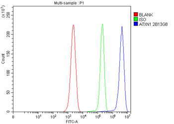
Flow Cytometry analysis of ANA-1 cells using anti-Ataxin 1 antibody. Overlay histogram showing ANA-1 cells (Blue line). To facilitate intracellular staining, cells were fixed with 4% paraformaldehyde and permeabilized with permeabilization buffer. The cells were blocked with 10% normal goat serum. And then incubated with mouse anti-Ataxin 1 Antibody (1 µg/1x10 6 cells) for 30 min at 20C. DyLight488 conjugated goat anti-mouse IgG (5-10 µg/1x10 6 cells) was used as secondary antibody for 30 minutes at 20C. Isotype control antibody (Green line) was mouse IgG (1 µg/1x10 6) used under the same conditions. Unlabelled sample without incubation with primary antibody and secondary antibody (Red line) was used as a blank control.
Flow Cytometry analysis of NRK cells using anti-Ataxin 1 antibody. Overlay histogram showing NRK cells (Blue line). To facilitate intracellular staining, cells were fixed with 4% paraformaldehyde and permeabilized with permeabilization buffer. The cells were blocked with 10% normal goat serum. And then incubated with mouse anti-Ataxin 1 Antibody (1 µg/1x10 6 cells) for 30 min at 20C. DyLight488 conjugated goat anti-mouse IgG (5-10 µg/1x10 6 cells) was used as secondary antibody for 30 minutes at 20C. Isotype control antibody (Green line) was mouse IgG (1 µg/1x10 6) used under the same conditions. Unlabelled sample without incubation with primary antibody and secondary antibody (Red line) was used as a blank control.
Flow Cytometry analysis of PC-3 cells using anti-Ataxin 1 antibody. Overlay histogram showing PC-3 cells (Blue line). To facilitate intracellular staining, cells were fixed with 4% paraformaldehyde and permeabilized with permeabilization buffer. The cells were blocked with 10% normal goat serum. And then incubated with mouse anti-Ataxin 1 Antibody (1 µg/1x10 6 cells) for 30 min at 20C. DyLight488 conjugated goat anti-mouse IgG (5-10 µg/1x10 6 cells) was used as secondary antibody for 30 minutes at 20C. Isotype control antibody (Green line) was mouse IgG (1 µg/1x10 6) used under the same conditions. Unlabelled sample without incubation with primary antibody and secondary antibody (Red line) was used as a blank control.
IHC analysis of Ataxin 1 using anti-Ataxin 1 antibody. Ataxin 1 was detected in a paraffin-embedded section of human hepatocellular carcinoma tissue. Heat mediated antigen retrieval was performed in EDTA buffer (pH8.0, epitope retrieval solution). The tissue section was blocked with 10% goat serum. The tissue section was then incubated with 2 µg/ml mouse anti-Ataxin 1 Antibody overnight at 4C. Peroxidase Conjugated Goat Anti-mouse IgG was used as secondary antibody and incubated for 30 minutes at 37C. The tissue section was developed using HRP Conjugated Mouse IgG Super Vision Assay Kit with DAB as the chromogen.
IHC analysis of Ataxin 1 using anti-Ataxin 1 antibody. Ataxin 1 was detected in a paraffin-embedded section of human laryngeal squamous cell carcinoma tissue. Heat mediated antigen retrieval was performed in EDTA buffer (pH8.0, epitope retrieval solution). The tissue section was blocked with 10% goat serum. The tissue section was then incubated with 2 µg/ml mouse anti-Ataxin 1 Antibody overnight at 4C. Peroxidase Conjugated Goat Anti-mouse IgG was used as secondary antibody and incubated for 30 minutes at 37C. The tissue section was developed using HRP Conjugated Mouse IgG Super Vision Assay Kit with DAB as the chromogen.
IHC analysis of Ataxin 1 using anti-Ataxin 1 antibody. Ataxin 1 was detected in a paraffin-embedded section of human thyroiditis tissue. Heat mediated antigen retrieval was performed in EDTA buffer (pH8.0, epitope retrieval solution). The tissue section was blocked with 10% goat serum. The tissue section was then incubated with 2 µg/ml mouse anti-Ataxin 1 Antibody overnight at 4C. Peroxidase Conjugated Goat Anti-mouse IgG was used as secondary antibody and incubated for 30 minutes at 37C. The tissue section was developed using HRP Conjugated M
* VAT and and shipping costs not included. Errors and price changes excepted