B7-H3 Antibody is supplied in PBS containing 0.02% sodium azide and 50% glycerol.
Form:
Liquid
Target:
CD276
Application Notes:
Application Notes: B7-H3 antibody can be used for detection of B7-H3 by Western blot at 0.25 µg/mL. Antibody can also be used for immunohistochemistry starting at 2 µg/mL and Immunocytochemistry starting at 1 µg/mL. For immunofluorescence start at 10 µg/mL. Flow cytometry at 1 µg/ml.Antibody validated: Western Blot in human samples, Immunohistochemistry in human samples, Immunocytochemistry in human samples, Immunofluorescence in human samples and Flow Cytometry in human samples. All other applications and species not yet tested
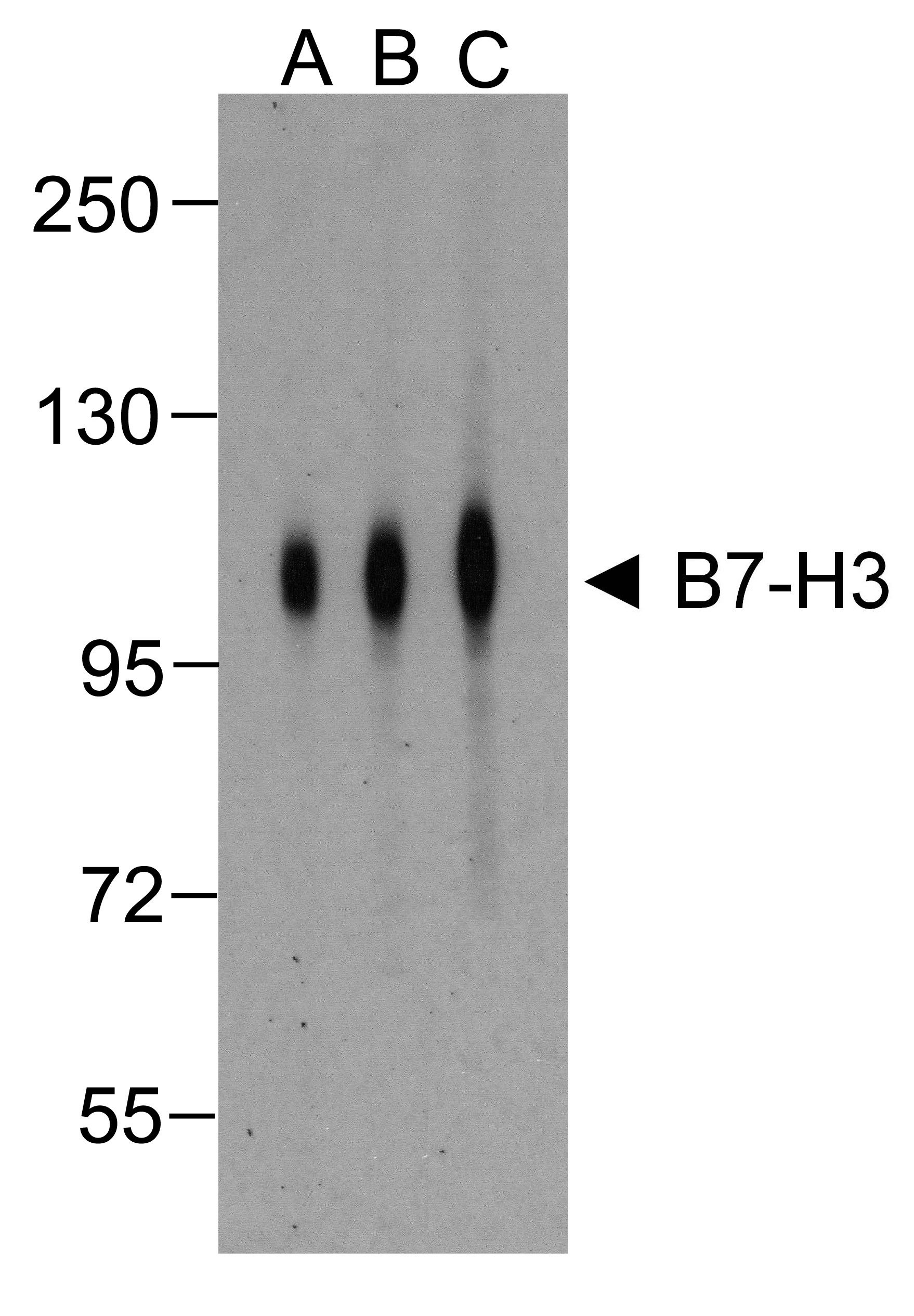
Western blot analysis of B7-H3 in HEK293 cells using B7-H3 antibody at (A) 0.25 (B) 0.5 and (C) 1 µg/ml.
Immunocytochemistry of B7-H3 in HEK293 cells using B7-H3 antibody and control mouse IgG antibody (left corner box) at 1 µg/ml.
Immunofluorescence of B7-H3 in HEK293 cells using B7-H3 Antibody at 5 µg/ml. Green: B7-H3 Antibody [2A7] (orb1239243) Blue: DAPI staining
Immunofluorescence of B7-H3 in human colon carcinoma tissue cells using B7-H3 Antibody at 10 µg/ml. Green: B7-H3 Antibody [2A7] (orb1239243) Blue: DAPI staining
Immunohistochemistry of B7-H3 in human colon carcinoma tissue using B7-H3 Antibody and control mouse IgG (corner box) at 2 µg/ml.
Flow cytometry analysis of B7-H3 in HEK293 cells using B7-H3 antibody at 1 µg/ml. Blue: untransfected HEK293 cells. Yellow: B7-H3 over expressing HEK293 cells.
Titration curve analysis of B7-H3 mAbs to detect recombinant B7-H3 in ELISA with orb1240155, orb1239243, orb1239244, orb1239247, and orb1239231 antibodies at decreasing concentrations.
* VAT and and shipping costs not included. Errors and price changes excepted