
BACE1 Antibody, Unconjugated, Rabbit, Polyclonal
Catalog Number:
BYT-ORB1239248
- Images (11)


| Article Name: | BACE1 Antibody, Unconjugated, Rabbit, Polyclonal |
| Biozol Catalog Number: | BYT-ORB1239248 |
| Supplier Catalog Number: | orb1239248 |
| Alternative Catalog Number: | BYT-ORB1239248-0.02,BYT-ORB1239248-0.1 |
| Manufacturer: | Biorbyt |
| Host: | Rabbit |
| Category: | Antikörper |
| Application: | ELISA, ICC, IF, IHC-P, WB |
| Species Reactivity: | Human, Mouse |
| Immunogen: | Anti-BACE antibody (orb1239248) was raised against a peptide corresponding to 17 amino acids near the carboxy terminus of human BACE. The immunogen is located within the last 50 amino acids of BACE. |
| Conjugation: | Unconjugated |
| Alternative Names: | BACE Antibody: ASP2, BACE, HSPC104, KIAA1149, Beta-secretase 1, Aspartyl protease 2, ASP2 |
| BACE1 Antibody |
| Application Notes: | Application Notes: WB: 1 µg/mL, IHC-P: 2.5 µg/mL, ICC: 10 µg/mL, IF: 20 µg/mL.Antibody validated: Western Blot in human and mouse samples, Immunohistochemistry, Immunocytochemistry and Immunofluorescence in mouse samples. All other applications and species not yet tested |
|
|
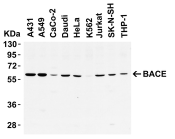
WB Validation in Human Cell Lines. Loading: 10 µg of lysate. Antibodies: BACE, orb1239248, 1 µg/mL, 1 h incubation at RT in 5% NFDM/TBST. Secondary: Goat Anti-Rabbit IgG HRP conjugate at 1:10000 dilution. |
|
|
KD Validation of BACE in DRG (Hyun, orb1239261). Decreased BACE1 expression in DRG following siRNA3 transfection. DRG neurons were transfected with 1 µg siRN |
|
|
|
|
|
Independent Antibody Validation (IAV) via Protein Expression Profile in Cell Lines. Loading: 15 µg of lysates per lane. Antibodies: BACE orb1239248 (1 µg/mL), BACE orb1273668 (1 µg/mL), beta-actin (1 µg/mL), and GAPDH (0.02 µg/mL), 1h incubation at RT in 5% NFDM/TBST. Secondary: Goat anti-rabbit IgG HRP conjugate at 1:10000 dilution. |
|
|
WB Validation in Mouse Tissues. Loading: 15 µg of lysate. Antibodies: BACE, orb1239248, 2 µg/mL, 1 h incubation at RT in 5% NFDM/TBST. Secondary: Goat Anti-Rabbit IgG HRP conjugate at 1:10000 dilution. |
|
|
Immunohistochemistry Validation of BACE in Human Liver. Immunohistochemical analysis of paraffin-embedded human liver tissue using anti-BACE antibody (orb1239248) at 2 µg/ml. Tissue was fixed with formaldehyde and blocked with 10% serum for 1 h at RT, antigen retrieval was by heat mediation with a citrate buffer (pH6). Samples were incubated with primary antibody overnight at 4C. A goat anti-rabbit IgG H&L (HRP) at 1/250 was used as secondary. Counter stained with Hematoxylin. |
|
|
Immunofluorescence Validation of BACE in Mouse Liver. Immunofluorescent analysis of 4% paraformaldehyde-fixed mouse liver tissue labeling BACE with orb1239248 at 10 µg/mL, followed by goat anti-rabbit IgG secondary antibody at 1/500 dilution (red) and DAPI staining (blue). |
|
|
KO and Overexpression Validation of BACE in Human and Mouse Brain and 293 Cells. Western blot analysis of the BACE1 (orb1239248) antibodys ability to recognize human and murine BACE1. The BACE1 antibody recognized both the mouse and human forms of BACE1. Lanes 1-4 are frontal cortex homogenates from human and mouse brains. Lane 1 is from a neurologically unimpaired aged human control case, lane 2 from a BACE1-deficient mouse, lane 3 from a nontransgenic mouse and lane 4 from hBACE1 transgenic mouse. Lanes 5-7 are lysates from HEK293T cells transfected with a plasmid vector expressing eGFP, mBACE1 and hBACE1, respectively. |
|
|
KD Validation of BACE in Mouse Brain. Characterization of the effects of lenti-siBACE1-6 expression in the brains of APP transgenic mice. (a-d) Anti-eGFP immunoreactivity in the hippocampus (the injection site) shows comparable and consistent expression of lenti-siRNA constructs in the dentate gyrus (dg) and stratus polymorphus (sp). (e) Anti-BACE1 immunoreactivity in the hippocampus of nontransgenic mice treated with lenti-siGlut4. (f) Reduced BACE1 immunostaining in the hippocampus of nontransgenic mice treated with lenti-siBACE1-6 vector. (g) Intense BACE1 immunoreactivity in the hippocampus of APP transgenic mice treated with lenti-siGlut4. (h) Reduced BACE1 expression in APP transgenic mice treated with lenti-siBACE1-6 vector. (i, j) Anti-BACE1 reacted with pyramidal cell bodies in the neocortex, which was not injected. |
|
|
KD Validation of BACE in Mouse Brain. Immunolabeling patterns of BACE1 expression and the lenti-siRNA distribution. Sections from APP transgenic mice treated with the eGFPtagged lenti siRNA vectors (green) were co-immunolabeled with an antibody against BACE1 (red) and imaged with the LSCM. All sections are from the hippocampus of treated mice. (a-c) Lenti-siBACE1-6-treated mice. Areas within the hippocampus expressing the eGFP tagged vector have reduced BACE1 immunolabeling. (d-f) Mice treated with the eGFP-tagged control lenti-siGlut4 show unchanged expression of BACE1 in the hippocampus. (g-i) Mice treated with a saline vehicle show unchanged expression of BACE1 in the hippocampus. |
|
|
KO Validation of BACE in MEF Cells. Wildtype and BACE -/- MEFs were exposed to HNE (15_M) for 2 h. BACE1 levels were examined by Western blot with anti-BACE antibodies (orb1239248). |
Product Guarantee and Expert Support
