
CDKN2A Antibody, Unconjugated, Rabbit, Polyclonal
Catalog Number:
BYT-ORB1239282
- Images (11)


| Article Name: | CDKN2A Antibody, Unconjugated, Rabbit, Polyclonal |
| Biozol Catalog Number: | BYT-ORB1239282 |
| Supplier Catalog Number: | orb1239282 |
| Alternative Catalog Number: | BYT-ORB1239282-0.02,BYT-ORB1239282-0.1 |
| Manufacturer: | Biorbyt |
| Host: | Rabbit |
| Category: | Antikörper |
| Application: | ELISA, IF, IHC-P, WB |
| Species Reactivity: | Human, Mouse, Rat |
| Immunogen: | Anti-CDKN2A antibody (orb1239282) was raised against a peptide corresponding to 18 amino acids near the amino terminus of human CDKN2A. The immunogen is located within the first 50 amino acids of CDKN2A. |
| Conjugation: | Unconjugated |
| Alternative Names: | CDKN2A Antibody: ARF, MLM, P14, P16, P19, CMM2, INK4, MTS1, TP16, CDK4I, CDKN2, INK4A, MTS-1, P19ARF, P16INK4, P16INK4A, P16-INK4A, Cyclin-dependent kinase 4 inhibitor A |
| CDKN2A Antibody |
|
|
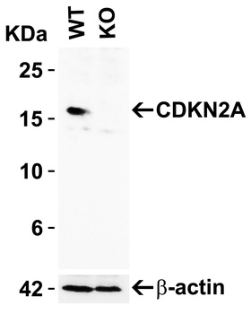
KO Validation of CDKN2A in 293 Cells. Loading: 10 µg of 293 WT cell lysates or CDKN2A KO cell lysates. Antibodies: CDKN2A, orb1239282 (2 µg/mL) and beta-actin, orb1240312 (1 µg/mL), 1 h incubation at RT in 5% NFDM/TBST. Secondary: Goat Anti-Rabbit IgG HRP conjugate at 1:10000 dilution. |
|
|
Immunohistochemistry Validation of CDKN2A in Human Colon Tissue. Immunohistochemical analysis of paraffin-embedded Human Colon Tissue using anti- CDKN2A antibody (orb1239282) at 10 µg/ml. Tissue was fixed with formaldehyde and blocked with 10% serum for 1 h at RT, antigen retrieval was by heat mediation with a citrate buffer (pH6). Samples were incubated with primary antibody overnight at 4C. A goat anti-rabbit IgG H&L (HRP) at 1/250 was used as secondary. Counter stained with Hematoxylin. |
|
|
Immunohistochemistry Validation of CDKN2A in Rat Colon Tissue. Immunohistochemical analysis of paraffin-embedded Rat Colon Tissue using anti- CDKN2A antibody (orb1239282) at 5 µg/ml. Tissue was fixed with formaldehyde and blocked with 10% serum for 1 h at RT, antigen retrieval was by heat mediation with a citrate buffer (pH6). Samples were incubated with primary antibody overnight at 4C. A goat anti-rabbit IgG H&L (HRP) at 1/250 was used as secondary. Counter stained with Hematoxylin. |
|
|
Independent Antibody Validation (IAV) via Protein Expression Profile in Cell Lines. Loading: 15 µg of lysates per lane. Antibodies: CDKN2A orb1239282 (4 µg/mL), CDKN2A orb1255478 (4 µg/mL), and beta-actin (1 µg/mL), overnight incubation at 4C in 5% NFDM/TBST. Secondary: Goat anti-rabbit IgG HRP conjugate at 1:10000 dilution. |
|
|
Recombinant Protein Test. Loading: 15 µg of lysates per lane. Antibodies: CDKN2A orb1239282 (2 µg/mL), overnight incubation at 4C in 5% NFDM/TBST. Secondary: Goat anti-rabbit IgG HRP conjugate at 1:10000 dilution. Lane 1: Human prostate (benign hyperplasia) Lane 2: Human CDKN2A recombinant protein (arrow: monomer and dimer). |
|
|
Western Blot Validation in Human Cell Lines. Loading: 15 µg of lysates per lane. Antibodies: CDKN2A, orb1239282 (2 µg/mL), 1h incubation at RT in 5% NFDM/TBST. Secondary: Goat anti-rabbit IgG HRP conjugate at 1:10000 dilution. |
|
|
Western Blot Validation in Human Normal and Cancer Tissue. Loading: 15 µg of lysates per lane. Antibodies CDKN2A orb1239282 (2 µg/mL), overnight incubation at 4C in 5% NFDM/TBST. Secondary: Goat anti-rabbit IgG HRP conjugate at 1:10000 dilution. Lane 1: Human breast, Lane 2: Human lung tumor, Lane 3: Human colon cancer. |
|
|
Western Blot Validation in Mouse Colon Tissue. Loading: 15 µg of lysates per lane. Antibodies: CDKN2A orb1239282 (A: 1 µg/mL, B: 2 µg/mL), 1h incubation at RT in 5% NFDM/TBST. Secondary: Goat anti-rabbit IgG HRP conjugate at 1:10000 dilution. |
|
|
Immunofluorescence Validation of CDKN2A in Human A431 Cells. Immunofluorescent analysis of 4% paraformaldehyde-fixed A431 cells labeling CDKN2A with orb1239282 at 20 µg/mL, followed by goat anti-rabbit IgG secondary antibody at 1/500 dilution (green) and DAPI staining (blue). |
|
|
Immunofluorescence Validation of CDKN2A in Human Colon Tissue. Immunofluorescent analysis of 4% paraformaldehyde-fixed human colon tissue labeling CDKN2A with orb1239282 at 20 µg/mL, followed by goat anti-rabbit IgG secondary antibody at 1/500 dilution (green) and DAPI staining (blue). |
|
|
Immunofluorescence Validation of CDKN2A in Rat Colon Tissue. Immunofluorescent analysis of 4% paraformaldehyde-fixed Rat Colon Tissue labeling CDKN2A with orb1239282 at 20 µg/mL, followed by goat anti-rabbit IgG secondary antibody at 1/500 dilution (red) and DAPI staining (blue). |
Product Guarantee and Expert Support
